
Frontonia tchibisovae Burkovsky, 1970
|
publication ID |
https://doi.org/ 10.5281/zenodo.180528 |
|
DOI |
https://doi.org/10.5281/zenodo.5661182 |
|
persistent identifier |
https://treatment.plazi.org/id/C44C3C09-FFCE-537F-FF1B-96B6DD2E49B5 |
|
treatment provided by |
Plazi |
|
scientific name |
Frontonia tchibisovae Burkovsky, 1970 |
| status |
|
Frontonia tchibisovae Burkovsky, 1970
( Figs. 7–9 View FIGURE 7 View FIGURE 8 View FIGURE 9 ; Table 1 View TABLE 1 )
Neither details of living morphology, nor a clearly-outlined diagnosis of this species was given ( Burkovsky 1970a). Hence a redescription and a redefined diagnosis based on both previous and present studies are supplied here.
Improved diagnosis: Marine Frontonia , in vivo ca. 130–250 × 80–190 µm, outline elliptical, dorsoventrally slightly flattened. 110–149 somatic, mostly 3 vestibular and 5–7 postoral kineties. Peniculus 1–3 each with 4 rows. One oval macronucleus. Single contractile vacuole centrally-located.
Sampling site: A scallop-larvae-rearing pond near Yantai, China, salinity ca. 27‰.
Voucher slides: One voucher slide with silver nitrate impregnated specimens (slide number: 2007:5:17:3) is deposited in the Natural History Museum, London, UK.
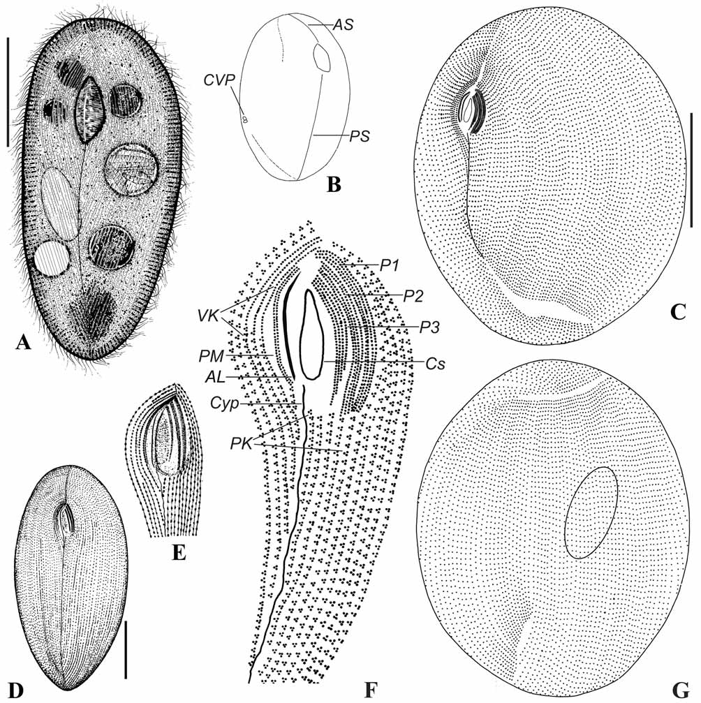
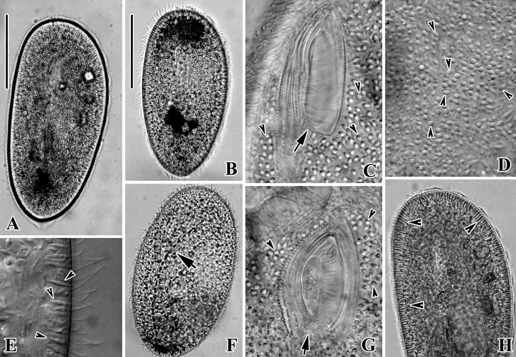
Description: Size highly variable, ca. 130–250 × 80–190 µm in vivo, but mostly about 200 × 140 µm. Body shape rather consistent, asymmetrical when viewed ventrally, slender elliptical in outline with narrowed posterior end; dorsoventrally slightly flattened ( Figs. 7 View FIGURE 7 A; 8A, 8B). Somatic cilia ca. 8 µm long. Buccal cavity triangular in shape, about 1/5–1/6 of body length, positioned in anterior 1/3 of cell length ( Figs. 7 View FIGURE 7 A; 8A, 8C, 8G; 9D). Cytoplasm hyaline and colourless, often with numerous tiny granules (<6 µm long), especially in frontal or caudal portion ( Figs. 7 View FIGURE 7 A; 8A, 8B). Macronucleus ellipsoid ( Fig. 7 View FIGURE 7 A). Extrusomes spindle-like, 5 µm long, densely distributing in the cortex ( Figs. 7 View FIGURE 7 A; 8A, 8C–E, 8G, 8H). Food vacuoles large, mainly containing algae and organic debris ( Figs. 7 View FIGURE 7 A; 8F).
Movement by rotating about the long axis of the cell, slightly thigmotactic, sometimes attaching to the bottom of the Petri dish and circling.
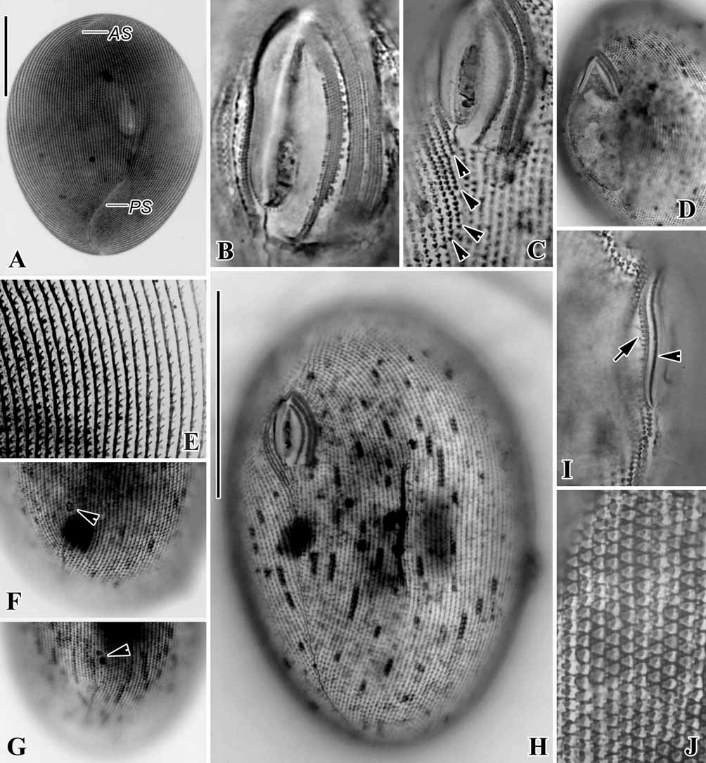
Infraciliature as shown in Figs. 7 View FIGURE 7 C, 7D, 7G and 9H. 127–149 somatic kineties ( Figs. 7 View FIGURE 7 C, 7G; 9E, 9H). Anterior and postoral sutures conspicuous ( Figs. 7 View FIGURE 7 B, 7C, 7G; 9A). 5–7 postoral kineties located posterior to the buccal apparatus and ending successively beside the cytopyge ( Fig. 7 View FIGURE 7 F).
Buccal apparatus typical of the genus. Mostly 3 vestibular kineties (at least two cells found with 4 VK, see Table 1 View TABLE 1 ) relatively short, starting from the anterior level of buccal cavity and ending before mid body level ( Figs. 7 View FIGURE 7 E, 7F; 9C, 9H). Peniculus 1 and 2 each with 4 (about equally long) rows of kinetosomes; P3 slightly shorter, also composed of 4 rows, with their length gradually lengthened from left to right ( Figs. 7 View FIGURE 7 F; 9B). Paroral membrane with an argentophilic line beside ( Figs. 7 View FIGURE 7 C, 7F; 9I).
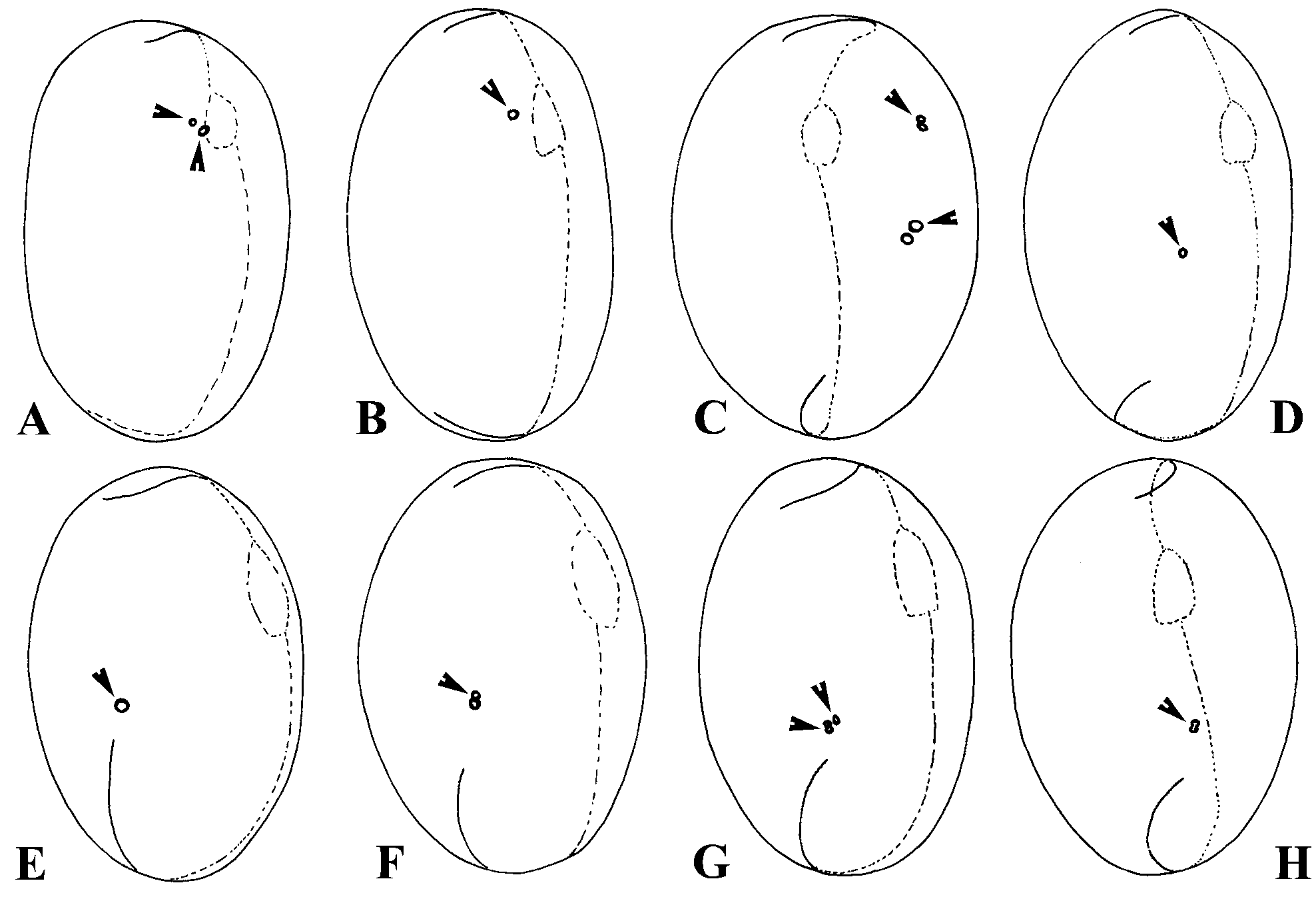
Silverline system typical of the genus ( Fig. 9 View FIGURE 9 J). 1–3, on average two CVP-like pore(s) right mid-dorsally located, near right margin of the cell (n=23; Figs. 2 View FIGURE 2 E–H; 9F, 9G).
Remarks: With reference to the morphology of living cells, size, especially the features of the somatic and buccal ciliature, the Qingdao isolate corresponds very well with the original report by Burkovsky (1970a) ( Fig. 7 View FIGURE 7 D, 7E). The only difference, as indicated originally, is the number of kineties in P3 (3 vs. 4 in the Qingdao population). This difference could be, in our opinion, a population-dependent diverse feature. Hence, we believe that the identification of our isolate is correct.
(B, D–F) and silver carbonate (C, G) impregnations. (A) Ventral view of a typical specimen. (B) To show the position of the CVP. (C, G) Ventral and dorsal sides of the infraciliature. (D, E) General view of the ventral side and the oral apparatus (after Burkovsky 1970a). (F) Detail of the oral apparatus, note the 3 vestibular kineties (VK), of which the anterior part consists of monokinetids. AL = argentophilic line; AS = anterior suture; Cs = cytostome; Cyp = cytopyge; P1–3 = peniculus 1–3; PK = postoral kinety; PM = paroral membrane; PS = postoral suture. Scale bars in (D) = 30 µm; in (A, C) = 60 µm.
The dorsally-located CVP-like pore(s) may probably derive from the CVP, as their location corresponds well with that of other known Frontonia species, or they are just structures, which were created by over staining.
With reference to the general morphology and the marine habitat, Frontonia tchibisovae is similar to the following morphospecies: F. elongata Burkovsky, 1970 , F. frigida Petz et al., 1995 , F. lynni , F. marina Fabre- Domergue, 1891 and. F. marisalbi Burkovsky, 1970 .
Frontonia elongata differs from F. t c h i b i s o v a e in having fewer somatic kineties (45–50 vs. 110–149 in the latter) and a smaller buccal cavity (1/14–1/11 of body length vs. 1/7–1/5) ( Fig. 10 View FIGURE 10 E, 10F; Table 2). F. m a r i n a has more vestibular kineties (6 vs. 3–4), kinety rows in P1 (6 vs. 4) and P2 (5 vs. 4), hence it can be clearly distinguished from F. tchibisovae ( Fig. 10 View FIGURE 10 K, 10L; Table 2).
Compared with Frontonia tchibisovae , F. frigida has a different cell shape (lanceolate with both ends conspicuously narrowed vs. elliptical with wide ends in F. t c h i b i s o v a e), more vestibular kineties (5 vs. 3–4 in F. tchibisovae ) and a smaller buccal cavity (1/10–1/7 vs. 1/7–1/ 5 in F. t c h i b i s o v a e) ( Fig. 10 View FIGURE 10 G, 10H; Table 2) ( Petz et al. 1995). F. marisalbi can be easily distinguished from F. t c h i b i s o v a e by its reniform body shape (vs. elliptical in the latter) and fewer rows in P3 (3 vs. 4 in the Qingdao population) ( Fig. 10 View FIGURE 10 M, 10N; Table 2) ( Burkovsky 1970a).
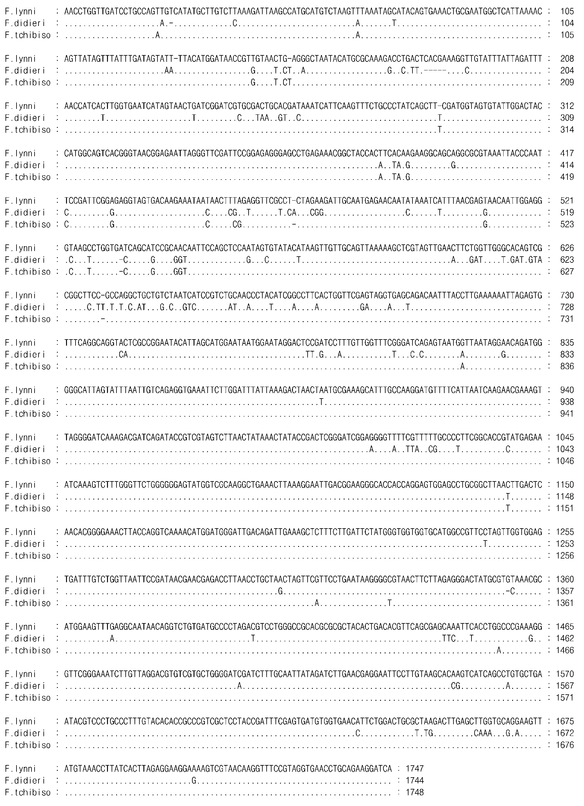
After rechecking the silver-nitrate impregnated specimens, it is confirmed that the P3 structure of Frontonia lynni was previously misinterpreted ( Long et al. 2005): there should be 4 kineties in P3 rather than 5 as described in the original report, of which the rightmost “kinety” is in fact not composed of kinetosomes, but argentophilic dots along the right-most kinety ( Fig. 10 View FIGURE 10 J, arrowhead). F. tchibisovae differs from F. l y n n i in having much more somatic kineties (110–149 vs. 71–83; Table 2). Besides, the dissimilarity of the two forms is firmly supported by 18S rRNA gene sequence data as the sequence of F. lynni differs in 30 nucleotides from that of F. t c h i b i s o v a e (structural similarity 98.3%) ( Fig. 11 View FIGURE 11 ).

No known copyright restrictions apply. See Agosti, D., Egloff, W., 2009. Taxonomic information exchange and copyright: the Plazi approach. BMC Research Notes 2009, 2:53 for further explanation.