
Enchytronia seongpanakiensis, Dózsa-Farkas & Felföldi & Nagy & Hong, 2018
|
publication ID |
https://doi.org/ 10.11646/zootaxa.4496.1.27 |
|
publication LSID |
lsid:zoobank.org:pub:7C536E1E-5D5A-4E2D-9E4F-28F3CEA9664C |
|
DOI |
https://doi.org/10.5281/zenodo.5950207 |
|
persistent identifier |
https://treatment.plazi.org/id/03D3D43A-E459-FFBF-2580-FD2FFB8CFB26 |
|
treatment provided by |
Plazi |
|
scientific name |
Enchytronia seongpanakiensis |
| status |
sp. nov. |
Enchytronia seongpanakiensis View in CoL sp. n.
( Figures 5G View FIGURE 5 , 10 View FIGURE 10 , 11 View FIGURE 11 )
Type material. Holotype: NIBRIV0000810592, slide No. 2252, adult, stained whole mounted specimen. Type locality: site 13, Mt. Hallasan , Seongpanak trail, Jeju Island, Korea, mixed-forest, N 33˚22'16.0", E 126˚33'51.6", 1352 m asl, 17.08.2016, leg. Y. Hong. Paratypes (in total 12 stained, adult specimens on slides and 9 specimens in 70% ethanol): NIBRIV0000810593, slide No. 2257, NIBRIV0000811384, slide No. 2364, from type locality. P.117.1.1–117.1.10, slide No. 2251–2256, 2363, 2365–2367 from type locality. In 70% ethanol: P117.2, from type locality 9 specimens .
Further material examined. 10 specimens investigated in vivo, 3 of them processed for DNA analysis.
Etymology. Named after the Seongpanak trail where it was found.
Diagnosis. The new species can be recognized by the following combination of characters: (1) small size (2.4– 4 mm in vivo), segments 22–25; (2) two chaetae per bundle but lateral chaetae absent in IX– XI; (3) large prostomial recess frontally; (4) clitellum in XII–XIII, only laterally; (5) brain incised posteriorly; (6) three pairs of preclitellar nephridia; (7) chloragocytes very large; (8) pharyngeal glands in IV and V united dorsally with ventral lobes in V, in VI free dorsally with large ventral lobes; (9) dorsal blood vessel from XII, blood light pink; anterior bifurcation in peristomium; (10) intestinal diverticula absent; (11) sperm funnels pear-shaped, 35–50 µm long in vivo; (12) male copulatory organs oval (26–32 µm long in vivo); (13) spermathecae attached to the oesophagus, ectal ducts 83–100 µm long, ampulla inconspicuous.
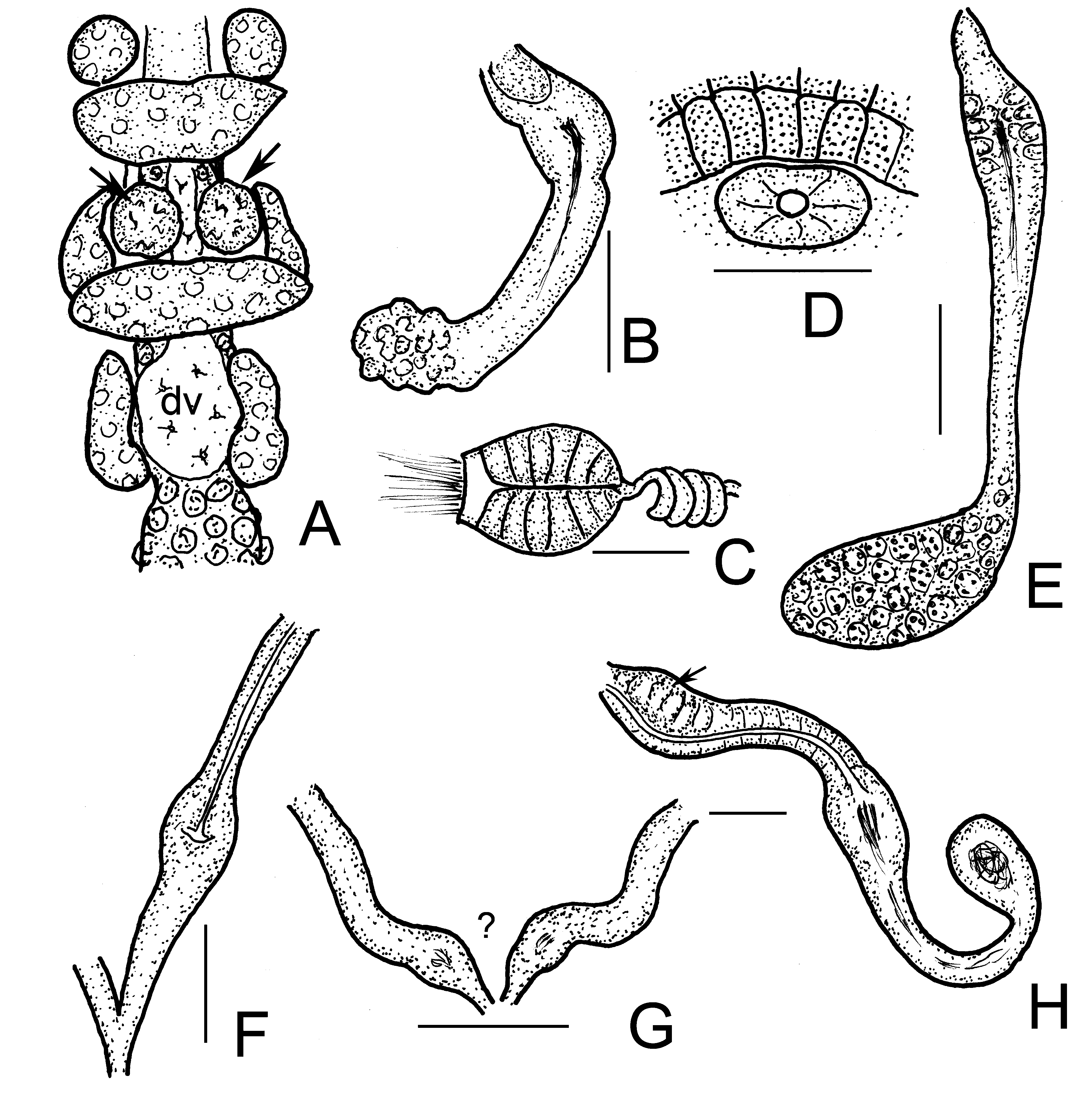
Description. Small worm. Holotype 3.15 mm long, 170 µm wide at VIII and 200 µm at clitellum (fixed), 23 segments. Length of paratypes 2.4–4.0 mm, width 175–220 µm at VIII and 21 0–240 µm at clitellum in vivo, length of fixed specimens 2.9–3.5 mm, width 130–170 µm at VIII and 140–200 µm at clitellum, segments 22–24. Chaetae straight with ental hook, ectally sharply pointed ( Fig. 10H View FIGURE 10 ), formula 2,0 – 2: 2 – 2. Laterally two chaetae from II– VIII, absent from IX preclitellarly, in XII absent laterally and also ventrally. Ventrally 2 chaetae per bundle; length ca. 23–28 µm, diameter ca. 2.5 µm; posteriorly increasing in size to 28–35 µm. (In one case, 3 and 4 chaetae were visible in ventral chaetal bundles at IV, V, probably replacement chaetae.) The frontal prostomial epithelium with a large vesicle-like recess at the frontal tip ( Fig. 10C View FIGURE 10 ). Head pore at 0/I ( Fig. 10C View FIGURE 10 ). Epidermal gland cells not seen. Body wall ca. 9–15 µm thick, preclitellarly thicker, cuticle inconspicuous <1 µm.
Brain about 80–100 µm long, 1.7–1.9 times longer than wide (fixed), incised posteriorly ( Figs. 10A–B View FIGURE 10 ). Postpharyngeal bulbs well developed ( Fig. 10J View FIGURE 10 ). Perikarya of II–IV fused into suboesophageal ganglion, segmental ganglia present from V on. Perikarya not only in the segmental ganglia ( Fig. 10D View FIGURE 10 ). Pharyngeal glands united in IV and V dorsally, primary ventral lobes in V; glands separate in VI, with large ventral lobe ( Figs. 10 I –J View FIGURE 10 ). Secondary ventral lobes absent. No oesophageal appendages, no intestinal diverticula. Chloragocytes with conspicuous vesicles from IV, dense layer from VII, very large cells ( Fig. 11A View FIGURE 11 ), diameter ca. 29–50 µm in vivo and 25–38 µm, fixed. Coelomocytes one type, mucocytes flat or spindle shaped in lateral view, slightly longer than wide, length ca. 26–36 µm in vivo (18–32 µm, fixed) filled with fine pale vesicles ( Fig. 10F–G View FIGURE 10 ). Gradual transition from oesophagus to intestine. Intestinal diverticula in VI not visible. Midgut pars tumida in XVII–XVIII, occupying 1–2 segment lengths, mostly not visible because of large chloragogen cells. Dorsal blood vessel from XII, blood light pink ( Fig. 11A View FIGURE 11 ). Anterior bifurcation in peristomium ( Fig. 11B View FIGURE 11 ). In some specimens, from the dorsal bifurcation a short branch is oriented forward ( Fig. 11C View FIGURE 11 ). Three pairs of preclitellar nephridia, at 7/8–9/10. Anteseptale with parts of nephridial body, no constriction at septum, postseptale slightly longer than anteseptale; tapers into short efferent duct, no terminal vesicles ( Figs. 10D–E View FIGURE 10 ). Clitellum in XII–XIII, only laterally developed ( Figs. 10 K–L View FIGURE 10 , 11E View FIGURE 11 ), i.e. absent dorsally and ventrally; cells in 27–32 separate rows. On either side between the granulocytes also hyalocytes in one or two longitudinal rows, which are not in contact with each other. The hyalocytes about 15–18 by 11–14 µm in size ( Fig. 10L View FIGURE 10 ). Seminal vesicle absent, spermatozoa ca. 25–37 µm long, heads 9–18 µm long in vivo (25–35 µm and 10–15 µm, respectively, fixed). Sperm funnel small, pear-shaped, 35–50 µm long in vivo (27– 40 µm, fixed) and 1.2–2.2 times longer than wide, collar conspicuous, about 5–10 µm high and slightly narrower than the funnel body ( Figs. 11F–H View FIGURE 11 ). Diameter of vas deferens ca. 5 µm. Male pore surrounded by small glands, 26– 32 µm long, 19–26 µm wide in vivo (17–26 µm long, 15–23 µm wide, fixed). Male pores apparently on body surface ( Fig. 11E View FIGURE 11 ), no bursa or bursal slits distinguished. No subneural glands. Spermathecae ( Figs. 5G View FIGURE 5 , 11J View FIGURE 11 ) proximally attached to the oesophagus, probably separately in V (the proximal part not visible because of large pharyngeal glands). Ectal ducts 46–65 µm long, 7–12 µm wide in vivo (35–45 µm long and 10–14 µm wide, fixed), no ectal gland, ampulla inconspicuous, ellipsoid mostly may not be distinctly set off from the ectal duct, if it is separable, diameter 8–12 µm wide in vivo (13–18 µm, fixed). Sperm not well visible. One or two large mature eggs at a time. In living specimens, often more unusual glandular sacculae visible on the ventral side ( Figs. 11I, K View FIGURE 11 ). We have not seen similar bodies in any other enchytraeid species. Unfortunately, they are not visible in fixed and stained specimens.
Distribution and habitat. In Korea, only in one sample (at site 13), mixed-forest in Seongpanak trail, Mt. Hallasan, 1352 m elevation.
Differential diagnosis. The new species differs from other Enchytronia species (except E. pygmaea Graefe & Schmelz, 2017 ) by the absence of the intestinal diverticula in VI, but we place it in the genus Enchytronia Nielsen & Christensen, 1959 because of the shape and arrangement of chaetae, absent laterally from IX to XI, by the posteriorly incised brain, the peristomial bifurcation of the dorsal blood vessel, the overall shape of the spermathecae, and only laterally developed clitellum and origin of the dorsal vessel at clitellum. Similarly, E. pygmaea did not have intestinal diverticula but it is clearly separate from Enchytronia seongpanakiensis sp. n. in the following main respects: smaller size (1.5–1.9 mm long, 17–19 segments vs. 2.4–4.0 mm, 22–25 segments), presence of only one lateral chaeta in postclitellar positions (vs. two chaetae), only one pair of preclitellar nephridia (vs. three pairs), and the posteriorly rounded brain (incised in the new species).


No known copyright restrictions apply. See Agosti, D., Egloff, W., 2009. Taxonomic information exchange and copyright: the Plazi approach. BMC Research Notes 2009, 2:53 for further explanation.