
Fridericia sousai, Schmelz, Rüdiger M. & Collado, Rut, 2013
|
publication ID |
https://doi.org/ 10.11646/zootaxa.3647.2.4 |
|
publication LSID |
lsid:zoobank.org:pub:33866E2B-6B0F-4124-A6A6-2B057E642149 |
|
DOI |
https://doi.org/10.5281/zenodo.5612038 |
|
persistent identifier |
https://treatment.plazi.org/id/301187BC-2C0D-FFF2-88B0-FDE34DDBF835 |
|
treatment provided by |
Plazi |
|
scientific name |
Fridericia sousai |
| status |
sp. nov. |
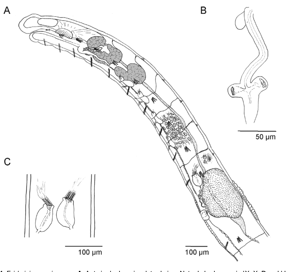
Fridericia sousai View in CoL sp. nov.
( Figs 1 View FIGURE 1 A–C, 5A, Table 3 View TABLE 3 )
Holotype. MNHML MB29-000297, adult spcm, stained whole mount. Portugal, Coimbra, in soil from the experimental field area of the Coimbra Higher School of Agriculture (ESAC), meadow site ( Table 2 View TABLE 2 ); II 2012.
Paratypes. Eight spms. MNHML MB29-000298 –301, stained whole mounts: 3 adults, 1 juvenile. ZMH OL 14518, fixed in Bouin's fluid, preserved in 70% ethanol: 1 adult, 1 juvenile. ZMH OL 14519, fixed in 70% ethanol, preserved in 100% ethanol: 2 spms.
Other material. 7 spms, investigated in vivo, preserved in collective sample vials, in the authors' collection.
Etymology. Named in honour of Paulo Sousa, soil zoologist, ecologist and ecotoxicologist at the University of Coimbra, research director of the TME experiments.
Diagnosis. Length <10 mm, less than 40 segments, max. 4 chaetae per bundle, often 3 chaetae per bundle, of same size in a bundle, clitellum girdle-shaped, present also between bursal slits, coelomo-mucocytes with refractile vesicles, nephridia absent at 10/11, chylus cells preclitellar with cell canals widened to lacunae, no seminal vesicle, sperm heads 20–25 μm, sperm funnel small, spermathecae separate entally, small ectal gland, two sessile diverticula without ciliated subchamber.
Description. Small Fridericia worms. Length 6–8 mm (viv), 4.5–6 mm (fix); diameter 0.15 mm (viv), 0.16 mm (fix), 0.2 mm at clitellum. Segment number 30–35. Chaetae max. 4 per bundle. Formula 2,3,4 – 4,3,2: 2,3,4 – 4,3,2. In preclitellar bundles mostly 4 chaetae, in postclitellar bundles 4 and 3 chaetae, more 3 than 4 in many specimens; two chaetae only in II and in the hindmost 1 or 2 segments. Anteriorly inner chaetae almost as large as outer, posteriorly all chaetae in a bundle of same size. Posterior chaetae slightly larger than anterior (52–55: 3–3.5 μm and c. 30–40: 2–3 μm, respectively). Chaetae increasing in size from II to VII (from c. 30 μm to c. 45 μm); chaetae in segments following clitellum as large as preclitellar; caudal chaetae largest (up to 60: 4 μm). Epidermal gland cells one row per segment, at chaetal level, few cells, one each dorsally of lateral chaetae. Body wall c. 15 μm thick, longitudinal muscle layer comparatively thin, 1–1.5x as thick as layer of ring muscles plus epidermis, cuticle very thin, barely visible at 400x magnification, estimated thickness <0.5 μm. Body surface slightly wavy in about 10 transverse rows per segment. All septa thin.
Brain rounded posteriorly, 80: 50 μm, sides almost parallel, anteriorly slightly convex, almost truncate. Pharyngeal glands dorsally connected in IV, V, separate in VI. Dorsal lobes all of same size, ventral lobes of same size in V and VI, small in IV. Oesophageal appendages short, unbranched. Chylus cells in IX–X, canals widened into elongate lacunae. Intestine widened in chylus region. Dorsal blood vessel from XIII. Midgut pars tumida from XX–XXIV, occupying 3–3.5 segments lengths. Preclitellar nephridia 4 pairs, from 6/7 to 9/10, length ratio anteseptale: postseptale about 1: 2; adseptal to medial rise of efferent duct, no terminal vesicle; postclitellar nephridia sparse, often unpaired, first from c. 17/18, shape as preclitellar, terminal rise of efferent duct. Coelomomucocytes with small refractile vesicles at periphery, cell length 15–20 μm, matrix almost hyaline (viv); lenticytes numerous but inconspicuous (viv), length 5–6 μm, i.e. about 1/3 of mucocyte length.
Clitellum girdle-shaped, cells in c. 25 dense rows, flat (i.e. cells wider than high), conspicuous in vivo, inconspicuous in whole mounts, also present between bursal slits; hyalocytes and granulocytes of same size, diameter 8–10 μm (fix). Seminal vesicle absent, developing sperm in anterior half of XI. Spermatozoa c. 80 μm long, heads 20–25 μm long. Sperm funnel small, length <1/2 body diameter, little longer than wide (e.g. 60: 52 μm, fix), with defined outline, not deformed, collar narrower than funnel body (32 μm). Vas deferens 4–5 μm wide (viv, fix). Male copulatory organ: bursal slit longitudinal; male gland small, only slightly projecting into body cavity, not flattened, c. 40 μm long, 30 μm wide, 30 μm high (fix). Subneural glands and other accessory glands absent. Spermatheca: small ectal gland, ectal duct shorter than body diameter, c. 1.5x as long as ampulla, c. 90 μm long, diameter c. 12 μm, proximal endpiece slightly projecting into ampulla, not widened; two broadly sessile diverticula or diverticula-like protrusions, diameter c. 18 μm, sperm-containing; ampulla tapering proximad, diverticula and ampulla apparently solid (viv) but lumina visible in whole mounts. One mature oocyte at a time, extending over 1.5–2 segment lengths.
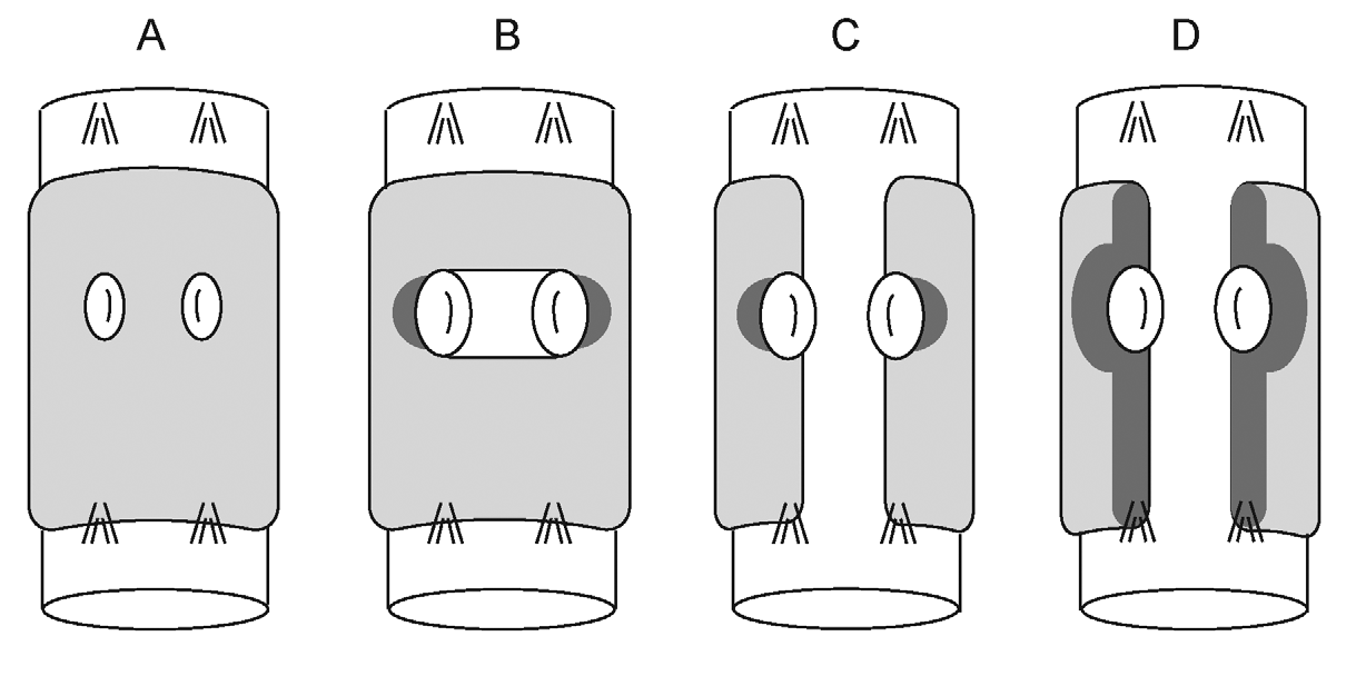
Remarks. Using the tabular comparison of Fridericia species with two spermathecal diverticula in Dózsa- Farkas (2009), F. sousai sp. nov. belongs to a group of 11 species, characterized by separate spermathecae and globular, hemispherical, or short-stalked diverticula. In this group, only F. isseli Rota, 1994 shares the following combination of characters with F. sousai : length <10 mm, less than 40 segments, max. 4 chaetae per bundle, oesophageal appendages short, unbranched, four pairs of preclitellar nephridia, chylus cells preclitellar, coelomocytes with refractile vesicles, seminal vesicle and subneural glands absent, spermatheca with ectal gland. F. isseli differs from the new species in the following traits, among others: (1) chylus cell canals branched, not widened (vs. unbranched, sac-like), (2) clitellum saddle-shaped (vs. girdle-shaped), (3) male gland tripartite with two small accessory glands (vs. accessory glands absent). In the key to Fridericia species in Schmelz (2003), F. sousai would key out together with F. isseli and with F. w a l d e n s t ro e m i Rota & Healy, 1999. The latter differs from F. s o u s a i in having 40–54 segments, five preclitellar pairs of nephridia, a small subneural gland in XIV, and proximally fused spermathecae with long-stalked diverticula, among other characters. Figure 5 View FIGURE 5 and Table 2 View TABLE 2 give a comparison of F. sousai with the other three new species of Fridericia from the same site.
No known copyright restrictions apply. See Agosti, D., Egloff, W., 2009. Taxonomic information exchange and copyright: the Plazi approach. BMC Research Notes 2009, 2:53 for further explanation.