
Hemienchytraeus cf. stephensoni Cognetti, 1927
|
publication ID |
https://doi.org/ 10.1080/00222933.2022.2140085 |
|
DOI |
https://doi.org/10.5281/zenodo.7426670 |
|
persistent identifier |
https://treatment.plazi.org/id/3A40CF59-FF80-FFF2-B26B-4527FC30DAA8 |
|
treatment provided by |
Plazi |
|
scientific name |
Hemienchytraeus cf. stephensoni Cognetti, 1927 |
| status |
|
Hemienchytraeus cf. stephensoni Cognetti, 1927 View in CoL
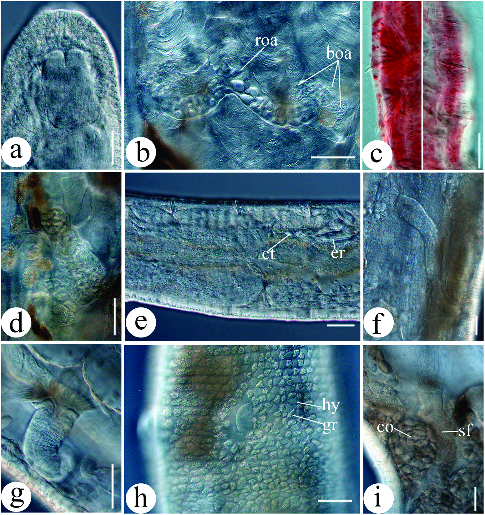
( Figure 18 View Figure 18 )
Hemienchytraeus stephensoni Cognetti, 1927 View in CoL . See Schmelz and Collado (2007) for list of synonymies, and for nomenclatural and taxonomic history of the nominal species.
Material investigated
GZO202007006, stained and whole-mounted, terminal segments absent, one mature specimen from site H; GZO202007007, stained and whole-mounted, one submature specimen from site H. CJJ 135, one mature specimen from site I, whole worm used for DNA extraction, preserved as total DNA.
Further material investigated
One mature specimen from site H, two mature specimens and one immature specimen from site I, six mature specimens from site 1, one mature specimen from site C and one mature specimen from site E, 12 in total, preserved in 75% ethanol.
Description
Small to medium-sized worms. Length 5.8–9.2 mm, diameter 0.21–0.34 mm at VII, 0.29– 0.35 mm at clitellum. Number of segments 32–38. Chaetae faintly sigmoid, anterior chaetae ca. 32.5–40 μm long and 3.75–5 μm thick, terminal chaetae enlarged (about 1.5× as large as anterior), ca. 52.5–62.5 μm long and 5–7.5 μm thick ( Figure 18c View Figure 18 ). Body wall 25.1–29.7 μm thick, cuticle very thin. Epidermal gland cells inconspicuous, cells nearly rectangular, 4–6 transverse rows per segment and arranged in regular order. Clitellum in XII–1/2XIII, girdle-shaped, well developed, granulocytes dense and arranged in reticulate with hyalocytes, hyalocytes isolated from each other, midventrally almost exclusively granulocytes ( Figure 18h View Figure 18 ).
Head pore on prostomium mid-dorsally. Brain nearly rectangular, incised anteriorly and slightly concave posteriorly, about 232 μm long and 153 μm wide (in vivo) ( Figure 18a View Figure 18 ). Oesophageal appendages arising mid-dorsally behind the pharynx in III, one root with large proximal chamber, two primary branches longer than root, with smaller branches; each primary branch bifurcating into three secondary branches, secondary branches thinner and shorter than primary branches ( Figure 18b View Figure 18 ). Three pairs of pharyngeal glands in IV–VI, united in IV–V and separated in VI dorsally, two pairs of secondary ventral lobes in V and VI, almost equal size. Dorsal vessel from XII to XIV, blood colourless. Four pairs of preclitellar nephridia from 6/7 to 9/10, anteseptale consisting of funnel and parts of nephridial body, ca. 43 μm long and 36 μm wide (in vivo); narrowed at septum, postseptale ca. 94 μm long and 59 μm wide (in vivo). Efferent duct originating from the middle of postseptale ( Figure 18d View Figure 18 ). Coelomocytes elliptic, brown in transmitted light, diameter 30– 33 μm (in vivo) ( Figure 18i View Figure 18 ).
Seminal vesicle absent. Sperm funnel tapering distad, collar indistinct, wider than funnel body, ca. 282–356 μm long and 50–57 μm wide at collar (in vivo) ( Figure 18f, g, i View Figure 18 ). Spermatozoa sparse, about 125 μm long (in vivo). Sperm duct elongate, with few coils in XIII. Male copulatory organ ( Figure 18h View Figure 18 ) small, male glandular body globular, diameter ca. 60 μm (in vivo). Spermathecae ( Figure 18e View Figure 18 ) free, not attached to oesophagus. Ectal pores at 4/5, no ectal gland. Ectal ducts muscular, short, ca. 61 μm long and 9 μm wide (in vivo); ampullae wider than ectal ducts (36 μm long, 14 μm wide, in vivo). Connecting tubes thinner than ampullae (337 μm long, 11 μm wide, in vivo), extending into VII, ending in ellipsoid ental reservoirs (70 μm long, 20 μm wide, in vivo). Ental reservoirs thin-walled, empty or with spermatozoa.
Remarks
Hemienchytraeus stephensoni was recorded in China by Xie et al. (1999), but the specimens were considered misidentified by the original authors, in a subsequent, detailed taxonomic revision of the species ( Schmelz and Collado 2007). The key characters (such as body size, chaetae, oesophageal appendage and spermatheca) of our specimens conform well to the description of H. stephensoni ( Schmelz and Collado 2007) .
No known copyright restrictions apply. See Agosti, D., Egloff, W., 2009. Taxonomic information exchange and copyright: the Plazi approach. BMC Research Notes 2009, 2:53 for further explanation.
|
Kingdom |
|
|
Phylum |
|
|
Class |
|
|
Order |
|
|
Family |
|
|
Genus |
Hemienchytraeus cf. stephensoni Cognetti, 1927
| Chen, Juanjuan, Schmelz, Rüdiger M., Zhang, Zuxu & Xie, Zhicai 2022 |
Hemienchytraeus stephensoni
| Cognetti 1927 |