
Membranacea spinata Qin & Zhang
|
publication ID |
https://doi.org/ 10.5281/zenodo.277935 |
|
DOI |
https://doi.org/10.5281/zenodo.6188425 |
|
persistent identifier |
https://treatment.plazi.org/id/03C6BD57-FF82-FFEF-FF5A-01D410F9FCC6 |
|
treatment provided by |
Plazi |
|
scientific name |
Membranacea spinata Qin & Zhang |
| status |
sp. nov. |
Membranacea spinata Qin & Zhang View in CoL , sp. n.
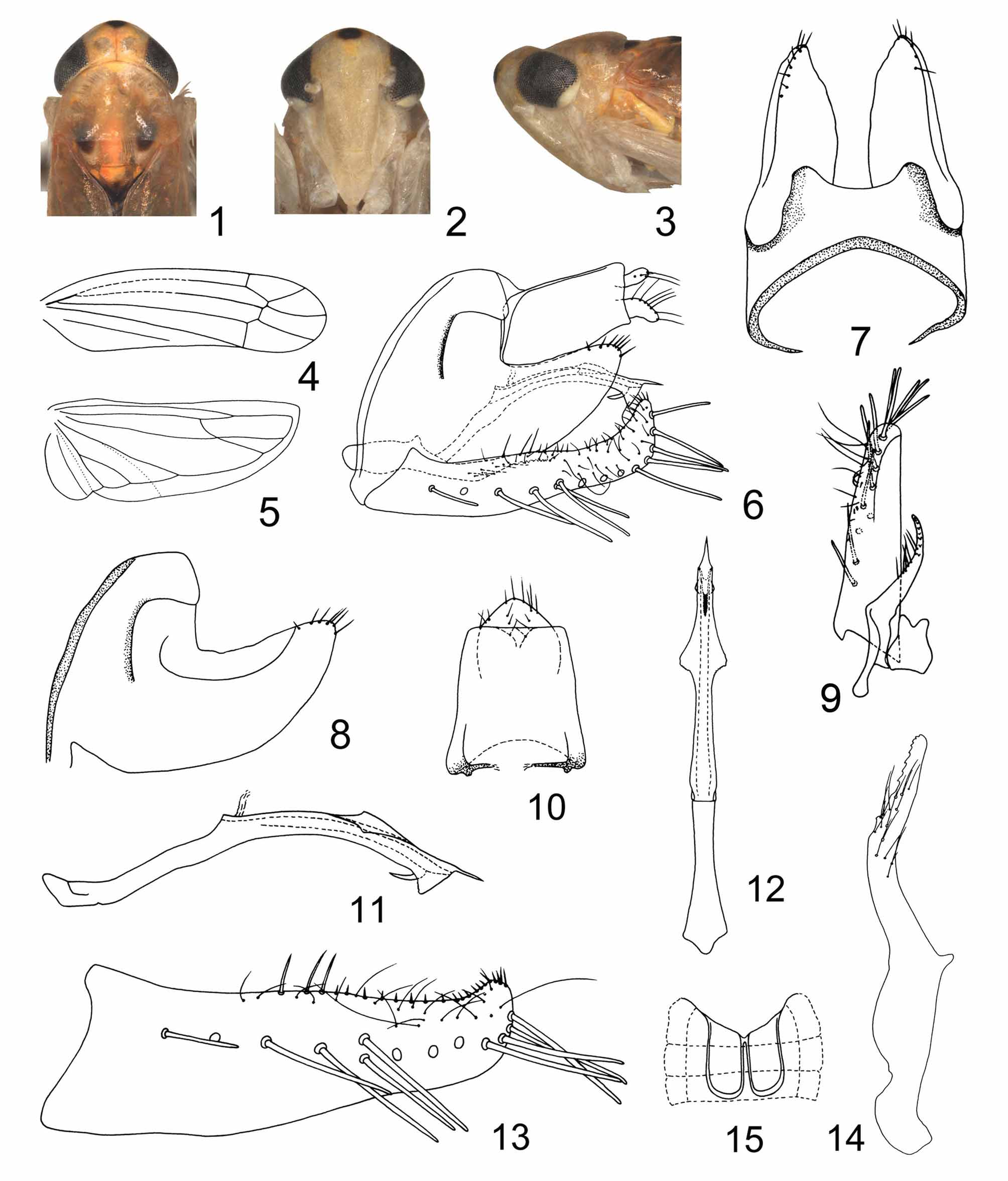
( Figs 1–15 View FIGURES 1 – 15 )
Type materials. Holotype, male ( NWAFU), Mt. Heng, Hunan Province, 11 Aug. 1985, coll. Yalin Zhang & Yonghui Chai. Paratypes, 3 males, same data as holotype; 1 male, 7 Aug. 1985, 2 males, 8 Aug. 1985, other data as holotype ( NWAFU).
Description. Length, male 4.0–4.3 mm.
General color reddish orange. Vertex centrally with a beige patch on each side of coronal suture, median black apical spot circled by creamy patch, the creamy patch extends basad of vertex which centrally joining the coronal suture ( Figs 1, 2 View FIGURES 1 – 15 ). Eyes dark. Ante- and postclypeus of face yellow. Genae beige. Pronotum reddish orange centrally and posteriorly, at anterior margin with an orange patch in middle and rest of arch of pronotum antero-laterally surrounded with irregular patches ( Fig. 1 View FIGURES 1 – 15 ). Centre of scutellum with a quadrate patch anteriorly, a semicircular patch caudad of scutoscutellar sulcus and two relatively small patch at each side of lateral margins, scutoscutellar sulcus brownish ( Fig. 1 View FIGURES 1 – 15 ). Forewing semi-transparent, reddish orange in basal 2/3 and yellowish in apical 1/3, hindwing hyaline, veins beige to brown. Abdomen blackish dorsally with irregular sordid yellow patches laterally on each tergite, beige ventrally. Legs yellow.
Basal sternal abdominal apodemes reaching end of segment V ( Fig. 15 View FIGURES 1 – 15 ). Male pygofer with ca. 7 rigid setae dorso-caudally ( Figs 6, 8 View FIGURES 1 – 15 ), dorsal bridge less than third of the total length of pygofer ( Fig. 7 View FIGURES 1 – 15 ), Subgenital plate broad in basal third, ventral margin curved dorsad in apical 2/3 leading to the plate slightly narrowing apicad, apex curved dorsad, slightly thickened dorsal margin in middle have 3 fairly long and broad setae forming the basal group, 13–14 lateral macrosetae, ca. 15 marginal microsetae and 2 irregular rows of feeble microsetae ( Figs 6, 13 View FIGURES 1 – 15 ). Paramere with 10 prominent teeth and ca. 14 long setae in cluster more cephald ( Fig. 14 View FIGURES 1 – 15 ). Aedeagal shaft with two pairs of flanges in dorsal aspect, the dorso-medial pair large and dentate basally at outer margins, another smaller pair near apex on ventral side, basad of these a spinous process medially, terminal part of shaft sharply narrowing to acuminated apex, preatrium slightly shorter than shaft, gonopore subterminal, ventral ( Figs 6, 11, 12 View FIGURES 1 – 15 ).
Remarks. Membranacea spinata Qin & Zhang , sp. n. differs from other members of this genus by the aedeagal shaft having a ventral spine near the apex.
Etymology. The species name is formed from “ spinatus ” (Latin; adjective), with the female termination –a, referring to the spine-like process of the aedeagus.
No known copyright restrictions apply. See Agosti, D., Egloff, W., 2009. Taxonomic information exchange and copyright: the Plazi approach. BMC Research Notes 2009, 2:53 for further explanation.