
Montezumellidae Ossó & Domínguez, 2013
|
publication ID |
https://doi.org/ 10.11646/zootaxa.4623.1.12 |
|
publication LSID |
lsid:zoobank.org:pub:0BA3A9D0-5191-42B4-99F9-C3C02830542C |
|
persistent identifier |
https://treatment.plazi.org/id/0398879E-FFF6-7055-FF49-FC47FACECEE3 |
|
treatment provided by |
Plazi |
|
scientific name |
Montezumellidae Ossó & Domínguez, 2013 |
| status |
|
Montezumellidae Ossó & Domínguez, 2013 View in CoL
Diagnosis (emended). Carapace from small/medium size ( Montezumella ) to large size ( Moianella ), from elongate to ovate subhexagonal shape, from slightly longer than wide to as wide as long, almost flattened; surface rugose, with coarse granules, and rows of granules forming short ridges of squamous aspect in the posterior half. Dorsal regions slightly swollen, well defined by shallow grooves, usually smooth. Hepatic region slightly swollen. Protogastric lobes swollen, anterior half of lobes both subdivided longitudinally by a median groove forming convergent ridges, that extend to the four frontal lobes or teeth; mesogastric region large, narrowing distally; metagastric region inflated; urogastric region narrow. Metagastric and cardiac regions well delimited transversally, and bounded laterally by a groove connecting the gastro-hepatic and cervical groove to the branchiocardiac groove. Cardiac region large. Intestinal region flat. Epibranchial lobes inflated. Mesobranchial and metabranchial regions slightly inflated. Front wide, bilobed, each lobe with two teeth or spines, axially divided by a notch. Orbits oblique, oval, complete; supra-orbital margin with three teeth separated by two fissures; serrated orbital corner continuous with the infraorbital margin which bears a prominent infra-orbital tooth.
Anterolateral margin short, straight to slightly convex, with four teeth or spines (excluding the outer orbital tooth) forward directed, subequal or decreasing in size. Posterolateral margin straight to convex, unarmed, stepped, edge decorated with tubercles or ridges; marked re-entrant of fifth pereiopod; branchiostegite not completely folded ventrally. Posterior margin from straight to slightly concave, short, rimmed.
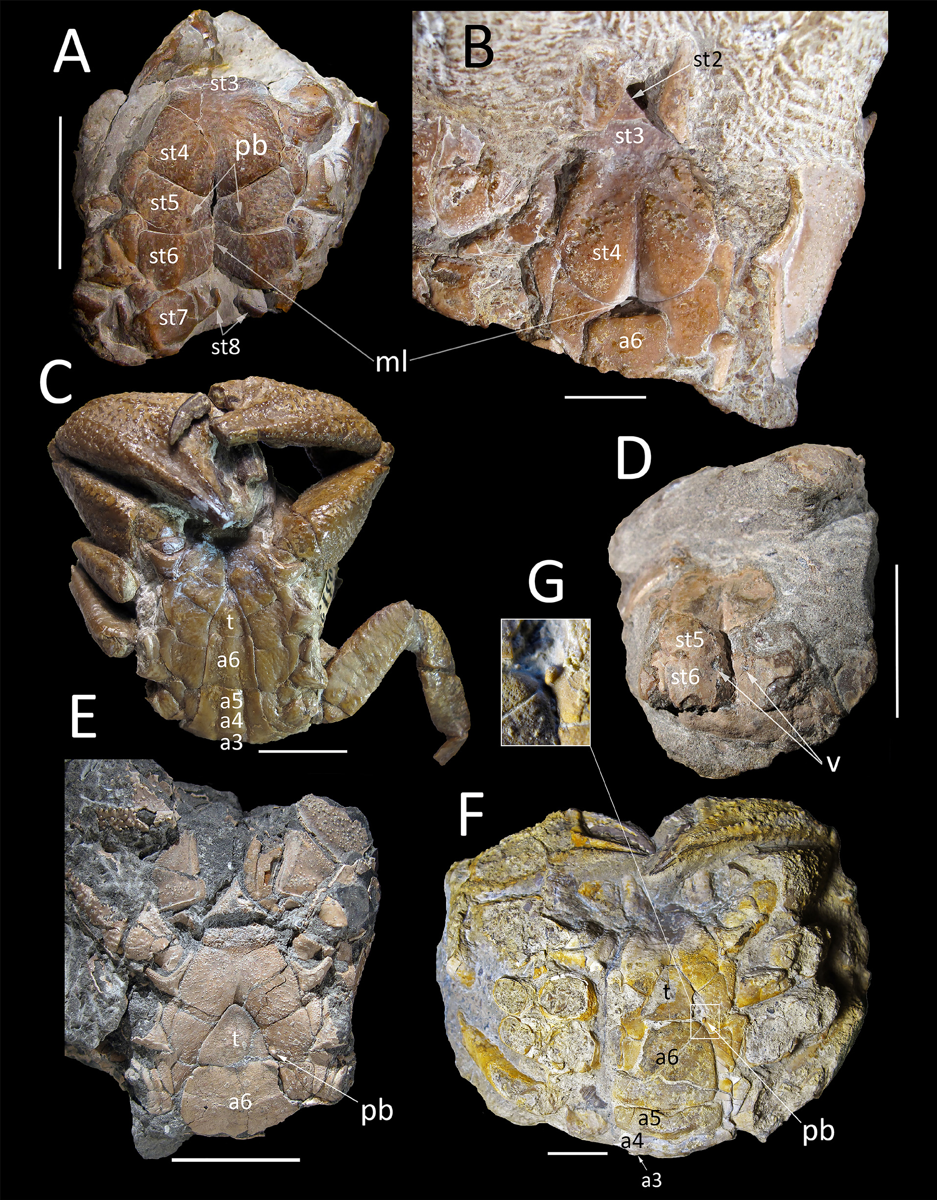
Antennular fossa deep, transverse, subtrapezoidal; basal article folded transversely. Basal article of antenna 2+3 subrectangular, elongate, directed obliquely between infraorbital tooth and antennular basal article. Proepistome thick, bluntly triangular. Ischium of third maxilliped with convex inner margin; merus subtrapezoidal. Male thoracic sternum narrow, elongate, metamerised. Suture 1/2 not recognizable, 3/4 only present laterally; sutures 2/3 and 4/5 to 7/8 complete; episternites well developed, delineated by complete sulci. Sternites 1 and 2 subtriangular; sternite 3 short, gynglyme (socket) receiving the coxo-sternal condyle of the mxp3 wide; sternite 4 elongate; sternites 5, 6, 7 subtrapezoidal. Median line present along the sternite 7 and extending until sternite 4. Press-button on sternite 5 close to suture 5/6. Sterno-pleonal cavity deep, narrow, V-shaped, reaching the half of sternite 4. Male pleon narrow, with all free somites and telson; somites 1, 2 not folded ventrally and in dorsal position; somites rectangular, somite 6 elongate. Female thoracic sternum larger than in males. Vulvae small, located inside deep sterno-pleonal cavity, on thoracic sternite 6 close to suture 5/6, completely covered by wide pleonal somite 6.
Chelipeds strong, slightly heterochelous in males and females. Merus robust, not fused with basis-ischium, subtriangular in section, inner surface concave, outer surface granulated and ridged. Carpus strong, massive, densely granulated, upper margin with two forward directed spines on inner and outer angles. Propodus strong, upper margin strongly tuberculated or spiny, outer margin convex, with two marked transverse spiny keels, the second one reaching the tip of pollex; dactylus curved, as long as propodus length, proximal third of upper margin spiny; both, pollex and dactylus almost homodontous, with central tooth of cutting edge slightly more developed. Ambulatory legs long, robust, sub-oval in section; dactylus long, acute lanceolate.
Remarks. The new specimen of Montezumella amenosi Vía, 1959 , which have well-preserved cephalic appendages, and a female of Moianella cervantesi with well-preserved thoracic and pleonal elements, allow and, at the same time, require a review of the family Montezumellidae .
Although Montezumellidae shares similar thoracic sternal pattern with Cancroidea (sensu Guinot et al. 2008; Schubart and Reuschel 2009), it cannot be retained in this superfamily in view of the clear differences, now confirmed by the data provided by the new specimens of Montezumella and Moianella . Hence, below we summarize the main differences after comparison with extant material.
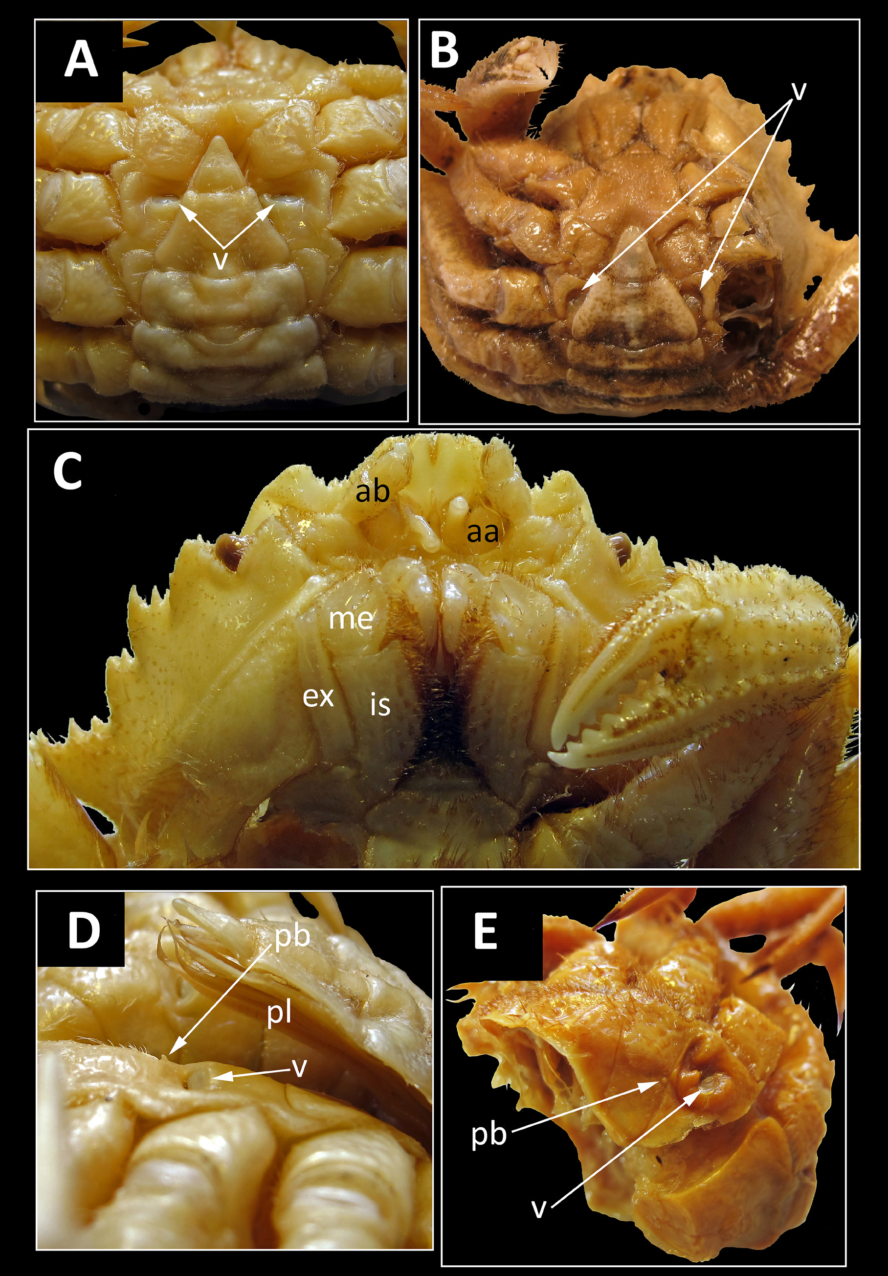
Members of Montezumellidae possess: a dorsal carapace slightly longer than wide, ornate with strong granules, tubercles and ridges, with regions well marked (vs. carapace wider than long in Cancridae ; ovate, and usually smooth in both families, Cancridae and Atelecyclidae ); only four anterolateral spines or teeth in Montezumellidae (vs. higher number of anterolateral teeth in Cancroidea); bilobed front, broader in Montezumellidae (vs. front narrow and uneven in Cancroidea); relatively broad and separated orbits in Montezumellidae (vs. small and close in Cancroidea) ( Fig. 3A, C View FIGURE 3 ; also see. Rathbun 1930b; Nations 1975; Schweitzer & Feldmann 2000); antennular fossae transversely subtrapezoidal, in which the antennules are folded transversely (vs. antennular fossae subquadrate and antennules folding longitudinally or slightly obliquely in Cancroidea); antennal basal article 2+3 narrower than basal (urinary) article (vs. much larger antennal basal article in Cancroidea); thick proepistome (vs. very narrow in Cancroidea) ( Fig. 3D, E View FIGURE 3 ; also see Rathbun 1930b: 176, pl. 87–88; Vía Boada et al. 1982: fig. 2d; Salva & Feldmann 2001; Spiridonov et al., 2014: fig. 2B; Davie et al. 2015a).
Which respect to thoracic sternal and pleonal features, differences are present as well. In Montezumellidae , the suture 2/3 is complete (vs. absent in Cancridae [ Fig. 3H View FIGURE 3 ], and well-marked but only laterally in Atelecyclidae [ Fig. 3G View FIGURE 3 ]); suture 3/4 is only marked laterally in Montezumellidae (vs. almost complete in Atelecyclidae ); the sternopleonal cavity is deep and reaches only half of sternite 4 in Montezumellidae (vs. sterno-pleonal cavity reaching sternite 3 in Cancroidea) ( Guinot 1979: pl. 9, figs. 4–7; Tavares & Cleva 2010: fig. 5E; Schram & Ng. 2012: fig. 3); vulvae are small in females of Montezumellidae (vs. larger in Cancroidea [ Fig. 3I View FIGURE 3 ]); pleonal somites 3, 4, 5 are free in males of Montezumellidae (vs. fused in Cancroidea [ Fig. 3G, H View FIGURE 3 ]); the pleonal somites 1, 2 are not folded ventrally, thus exposed in dorsal position, in Montezumellidae (vs. ventrally folded in Cancridae [e.g.: Rathbun 1930b; Bonfiglio & Donadeo 1982] and in Atelecyclidae [ Tavares & Cleva 2010, fig. 3A; Fig. 3B, C View FIGURE 3 ]) (e.g. Rathbun 1930b; Guinot 1979; Schweitzer & Feldmann 2000; Schram & Ng 2012; Davie et al. 2015a).
The morphological differences cited here, therefore are deemed enough to remove Montezumellidae from Cancroidea and raise it to superfamily rank.
The differences between Montezumelloidea n. superfam. and apparently related taxa, are summarized herein, making emphasis on the new characters now available, such as the cephalic appendages. Formerly, in introducing Montezumellidae as a new family, Ossó & Domínguez (2013), largely discussed the traits shared with closest relatives and listed its clearly distinguishable characters. Montezumelloidea n. superfam. shares with the families and genera grouped by Guinot (1979: 81–86, table 1), higher level characters such as the thoracic sternum with complete sutures 4/5 to 7/8 (see Ossó & Domínguez 2013: 289–293, and references therein).
An array of differences amongst Cheiragonidae (Cheiragonoidea Ortmann, 1893 [sensu Ng et al. 2008]) ( Fig. 4 View FIGURE 4 ), Montezumella , and Montezumellidae , was already listed by Ossó & Domínguez (2013: 289–292). In any case, the most distinctive character that excludes members of Montezumelloidea n. superfam. from Cheiragonoidea, is that the female of Cheiragonoidea possess exposed vulvae located laterally on the sternite 6 ( Fig. 4A, B View FIGURE 4 ; also Benedict 1892: 223, pl. 26, figs. 3, 6; Guinot 1979: 178, pl. 25, figs. 1–3; 1993: 1232; Števčić 1988; Schweitzer & Salva, 2000: 285; Karasawa & Schweitzer 2004: 144; Guinot et al. 2013: 28, fig. 3). By contrast, in Montezumelloidea n. superfam., the vulvae are located inside the sterno-pleonal cavity and are completely covered by the pleon, as usual in Eubrachyura ( Fig. 2D, E, F View FIGURE 2 ; also Ossó & Domínguez 2013: fig. 5 b, b’, c–e). In Montezumelloidea n. superfam., the somites 1, 2 are in dorsal position, whereas in Cheiragonidae the pleon appears only partially exposed dorsally, with only the first somite appearing in dorsal view (at least the somite 1 plus the entire somite 2 or only a part), thus the pleon is also incompletely folded, but to a lesser extent (see Benedict 1892: pls. 25–27). Furthermore, the new available specimens show the antennal basal article narrower than the preceding urinary article, and antennae 2+ 3 clearly divergent ( Fig. 1C, D View FIGURE 1 ), in contrast to species of Cheiragonoidea where antennae 2+3 are stout and convergent, respectively ( Fig. 4C View FIGURE 4 ). In Montezumelloidea n. superfam., the exopod of the third maxilliped is longer than the merus, whereas in Cheiragonoidea it is not as long as the merus ( Figs. 1C, D View FIGURE 1 , 4C View FIGURE 4 ); additionally, the proepistome is larger than in Cheiragonoidea ( Figs. 1 View FIGURE 1 C–D, 4C; cf. Benedict 1892, pl. 26, figs. 2, 5; Kamio et al. 2000, fig. 1). In adult females of Erimacrus isenbecki ( Brandt, 1848) and Telmessus cheiragonus ( Tilesius, 1812) , the press button is present but vestigial in mature females (vs. present and well-developed in adult females of Montezumelloidea n. superfam. ( Fig. 2E, F, G View FIGURE 2 ; Guinot, 1979: 137, pl. 25, figs 1–3; Guinot & Bouchard 1998). Reexamination of extant material in the MNHN collections, revealed that females of Erimacrus isenbecki present a small press button near the suture 5/6, not covered by the pleon, and thus exposed ( Fig. 4D View FIGURE 4 ). Likewise, females of Telmessus acutidens ( Stimpson 1858) and T. cheiragonus possess a small vestigial press button ( Fig. 4B, E View FIGURE 4 ). However, they are not functional due to the presence of pleopods and the lack of the socket on the corresponding part of the pleon (somite 6), as is often the case in mature female brachyurans. As previously stated by Ossó & Domínguez (2013: 289, 292, 296), according to the available fossil record, we do not recognize any fossil representative of Cheiragonoidea.
An extensive and detailed comparison between Montezumellidae and other taxa besides Cancroidea and Cheiragonoidea, but with which it also shares similar thoracic structures, e.g., a narrow and completely metamerised sternum and similar dorsal features, was carried out by Ossó & Domínguez (2013: 292, 293). These comparisons, which support a clear distinction between these taxa, are also valid for Montezumelloidea n. superfam. and are not be repeated herein. Nevertheless, a comparison taking into account the sterno-abdominal features and cephalic appendages, preserved in the new specimens, is summarized below.
In Corystes cassivelaunus ( Pennant, 1777) View in CoL (Fig. 5A, B) type species of Corystes Bosc, 1802 View in CoL , the type genus of Corystoidea Samouelle, 1819 (sensu Ng et al., 2008 [see Davie et al. 2015b: 945]), the antennules are folded longitudinally (vs. transversely in Montezumelloidea n. superfam.), and a pleonal-locking mechanism is lacking (vs. present in Montezumelloidea n. superfam.); the first pleonal somites is visible dorsally, as in Montezumelloidea n. superfam. (also Guinot 1979: 132; Ng. et al. 2008: fig. 39; Davie et al. 2015a: 1075). In Trichopeltarion nobile A. Milne-Edwards, 1880 View in CoL (Fig. 5C), type species of Trichopeltarion A. Milne-Edwards, 1880 View in CoL , the type genus of Trichopeltarioidea Tavares & Cleva, 2010, the antennulae are folded longitudinally (vs. transversely in Montezumelloidea n. superfam.) the basal article of antennae 2+3 rises convergently to the axis (vs. divergently to the axis in Montezumelloidea n. superfam.). Additionally, the limit between the endostome and epistome is poorly defined (vs. well defined in Montezumelloidea n. superfam.). The first pleonal somite is visible dorsally, as in Montezumelloidea n. superfam. (see Tavares & Cleva 2010). In Kraussia rugulosa ( Krauss, 1843) View in CoL (Fig. 5D, F), type species of Kraussia Dana, 1852 View in CoL , the type genus of Kraussiinae Ng, 1993 View in CoL ( Xanthidae MacLeay, 1838 View in CoL ), the antennules are folded obliquely (vs. transversely in Montezumelloidea n. superfam.); the antennae 2+3 rise longitudinally directed (vs. obliquely divergent in Montezumelloidea n. superfam.); the pleonal somites 1, 2 appear not folded ventrally and are in dorsal position, as in Montezumelloidea n. superfam. (Figs.5D, F; also Ng 1993). In Pilumnoides perlatus ( Poeppig, 1836) View in CoL (Fig. 5G, I), type species of Pilumnoides Lucas in H. Milne Edwards & Lucas, 1844 View in CoL , the type genus of Pilumnoididae Guinot & Macpherson, 1987 View in CoL , the antennules are folded nearly transversely (Fig. 5H; also Davie et al. 2015a: 1106;), as in Montezumelloidea n. superfam.; however, in Montezumelloidea n. superfam., the somites 1, 2 are not folded ventrally and in dorsal position, whereas in P. perlatus View in CoL they are folded ventrally (Fig. 5G; also Guinot & Macpherson 1987: pl. 1, fig. A). In any case, the differences highlighted by Ossó & Domínguez (2013: 292, 293) precludes any close relationship among both taxa, and likewise with all the taxa listed therein.


No known copyright restrictions apply. See Agosti, D., Egloff, W., 2009. Taxonomic information exchange and copyright: the Plazi approach. BMC Research Notes 2009, 2:53 for further explanation.
|
Kingdom |
|
|
Phylum |
|
|
Class |
|
|
Order |
|
|
Family |
Montezumellidae Ossó & Domínguez, 2013
| Ossó, Àlex & Domínguez, José Luis 2019 |
Kraussiinae
| Ng 1993 |
Pilumnoididae
| Guinot & Macpherson 1987 |
Trichopeltarion nobile
| A. Milne-Edwards 1880 |
Trichopeltarion
| A. Milne-Edwards 1880 |
Kraussia
| Dana 1852 |
Pilumnoides
| Lucas in H. Milne Edwards & Lucas 1844 |
| Krauss 1843 |
Xanthidae
| MacLeay 1838 |
Pilumnoides perlatus (
| Poeppig 1836 |
Corystes
| Bosc 1802 |
Corystes cassivelaunus (
| Pennant 1777 |