
Orbiniella petersenae, Parapar, Julio, Moreira, Juan & Helgason, Gudmundur Vidir, 2015
|
publication ID |
https://doi.org/ 10.11646/zootaxa.4006.2.5 |
|
publication LSID |
lsid:zoobank.org:pub:C6FF2AFA-3BD7-4B22-BE72-C48984714099 |
|
DOI |
https://doi.org/10.5281/zenodo.5661910 |
|
persistent identifier |
https://treatment.plazi.org/id/C268879D-FF84-723C-E0AA-E975FBB5E06B |
|
treatment provided by |
Plazi |
|
scientific name |
Orbiniella petersenae |
| status |
sp. nov. |
Orbiniella petersenae View in CoL sp. nov.
( Figs. 1 View FIGURE 1 , 3–9 View FIGURE 3 View FIGURE 4 View FIGURE 5 View FIGURE 6 View FIGURE 7 View FIGURE 8 View FIGURE 9 )
Material examined. A total of 222 specimens of Orbiniella petersenae sp. nov. were collected. Holotype: IINH 35670 (BIOICE sample 3636; 1,844−1,849 m depth, incomplete specimen). Paratypes: IINH 29795 (BIOICE sample 2430; 1,007−1,016 m; 26 spec.); IINH 34892 (BIOICE sample 2444; 133−148 m; 2 spec.); IINH 34894 (BIOICE sample 3611; 188−197 m; 16 spec. EtOH fixed); IINH 35669 (BIOICE sample 3632; 6 specimens on SEM stub); IINH 35671 ( BIOCE sample 3636; 1,844−1,849 m; 94 spec.); IINH 35672 ( BIOCE sample 3636; 1 posterior end;); MNCN 16.01/16549 (BIOICE sample 3636; 19 paratypes); IINH 29822 (BIOICE sample 3640; 1,908−1,915 m; 46 spec.); IINH 34897 (BIOICE sample 3640; 1 posterior end on SEM stub); IINH 35673 (BIOICE sample 3640; 3 specimens imaged with µCT); IINH 34899 (BIOICE sample 3656; 1,490−1,492 m; 9 spec.; EtOH fixed).
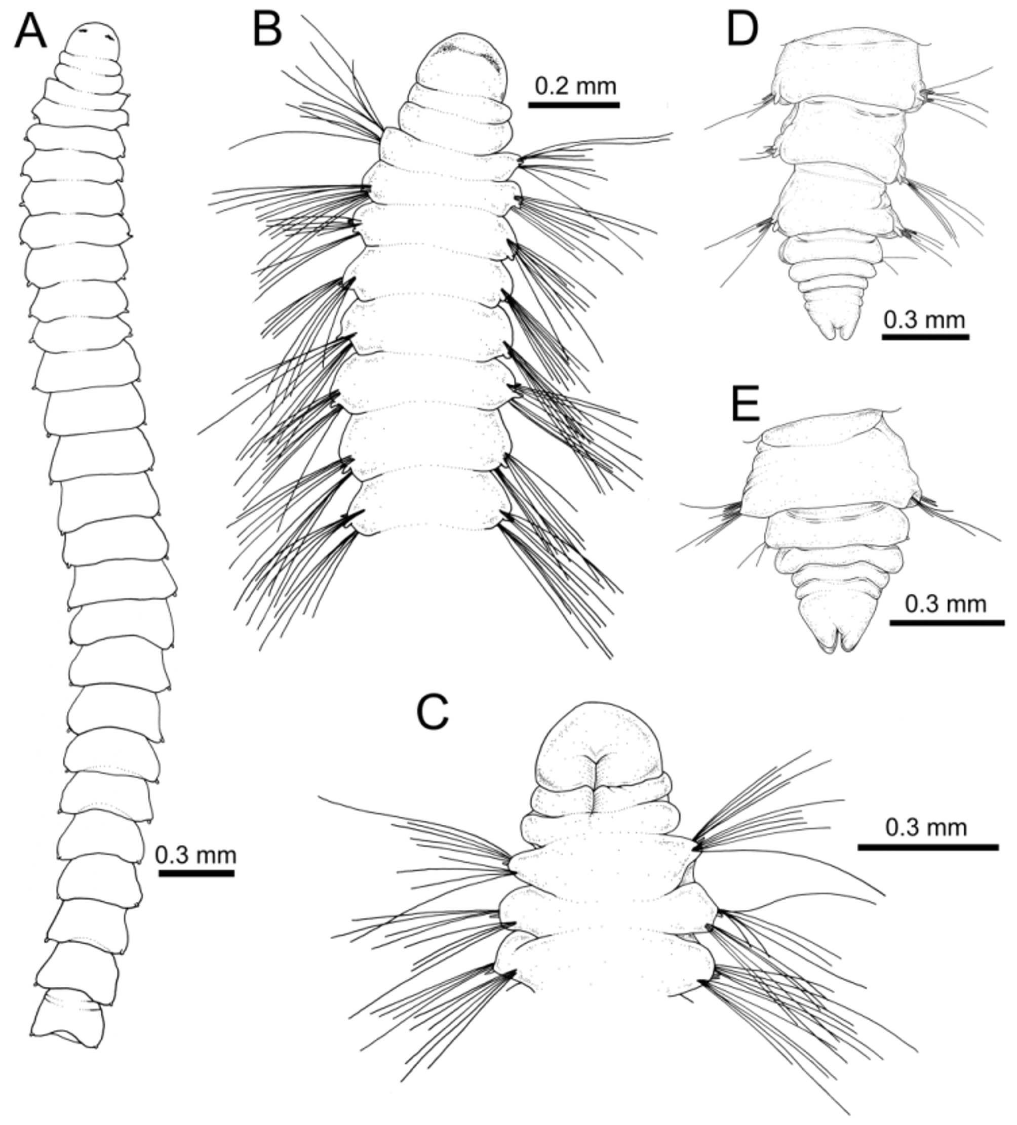
Description of holotype. Anterior fragment, body of uniform width, 6.0 mm long, 0.3 mm wide at level of first chaetiger, with 25 chaetigers; no body external regionalization ( Fig. 3 View FIGURE 3 A), lacking pigmentation. Prostomium rounded, wider than long ( Fig. 3 View FIGURE 3 B–C), provided with two dorsal brownish semi-lunar patches (diffuse photoreception area with ocelli?). No conspicuous nuchal organs observed. Two achaetous peristomial annuli, first narrower than second, distinctly separated from first chaetiger ( Fig. 3 View FIGURE 3 B–C). From CH 9 onwards segments becoming longer, more square-shaped ( Fig. 3 View FIGURE 3 A). Segment annulation not clear under light microscope (but see SEM observations of paratypes below).
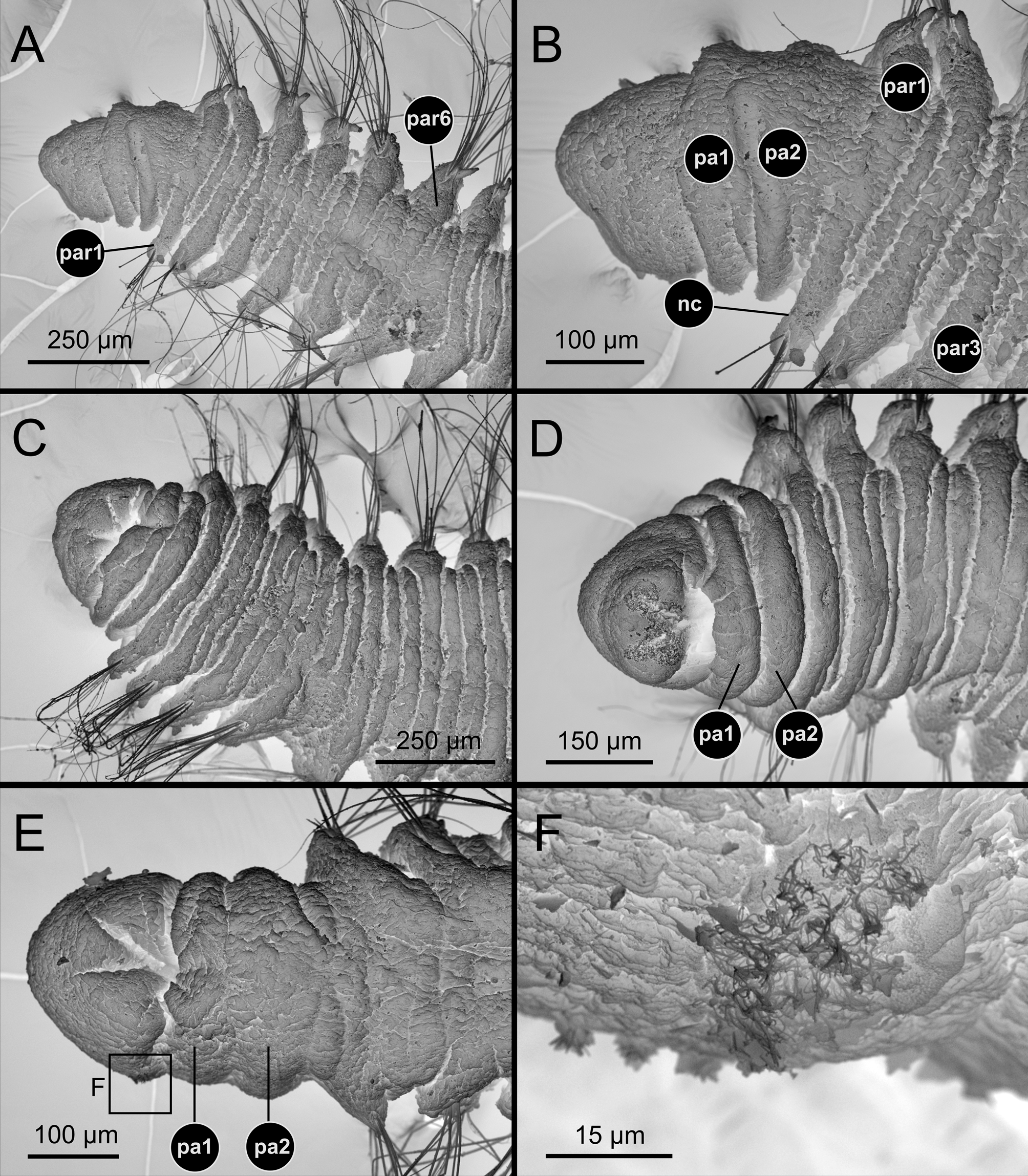
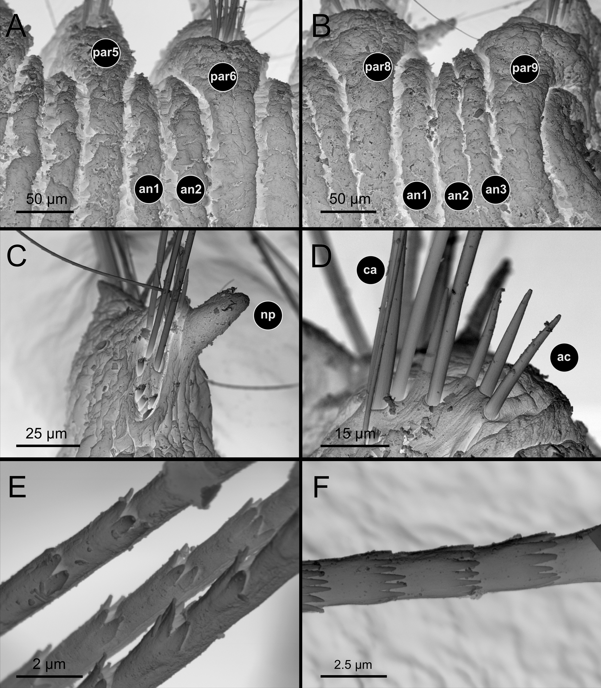
Notopodia with small, postchaetal papilla present from CH 1, sometimes located in between the two parapodial rami; papilla absent in neuropodia ( Fig. 3 View FIGURE 3 B and also see SEM micrographs of paratypes: Figs 4 View FIGURE 4 A, 5C, 6C, E). Notopodia and neuropodia bearing each 7–10 long crenulated capillaries and 1–3 short, stout smooth aciculae ( Fig. 3 View FIGURE 3 C and also see SEM micrographs of paratypes: Figs 5 View FIGURE 5 C–F, 6C, E); capillary chaetae with crenulation occurring on one side along half length. Capillaries in first segments longer than body width ( Fig. 3 View FIGURE 3 B).
Variation. The study of paratypes under stereomicroscope and SEM allowed elucidating the structure and range of variability of some body features. The species shows great body fragility; despite the high number of specimens obtained, none was complete. Longer anterior fragments are of about 23 chaetigers.
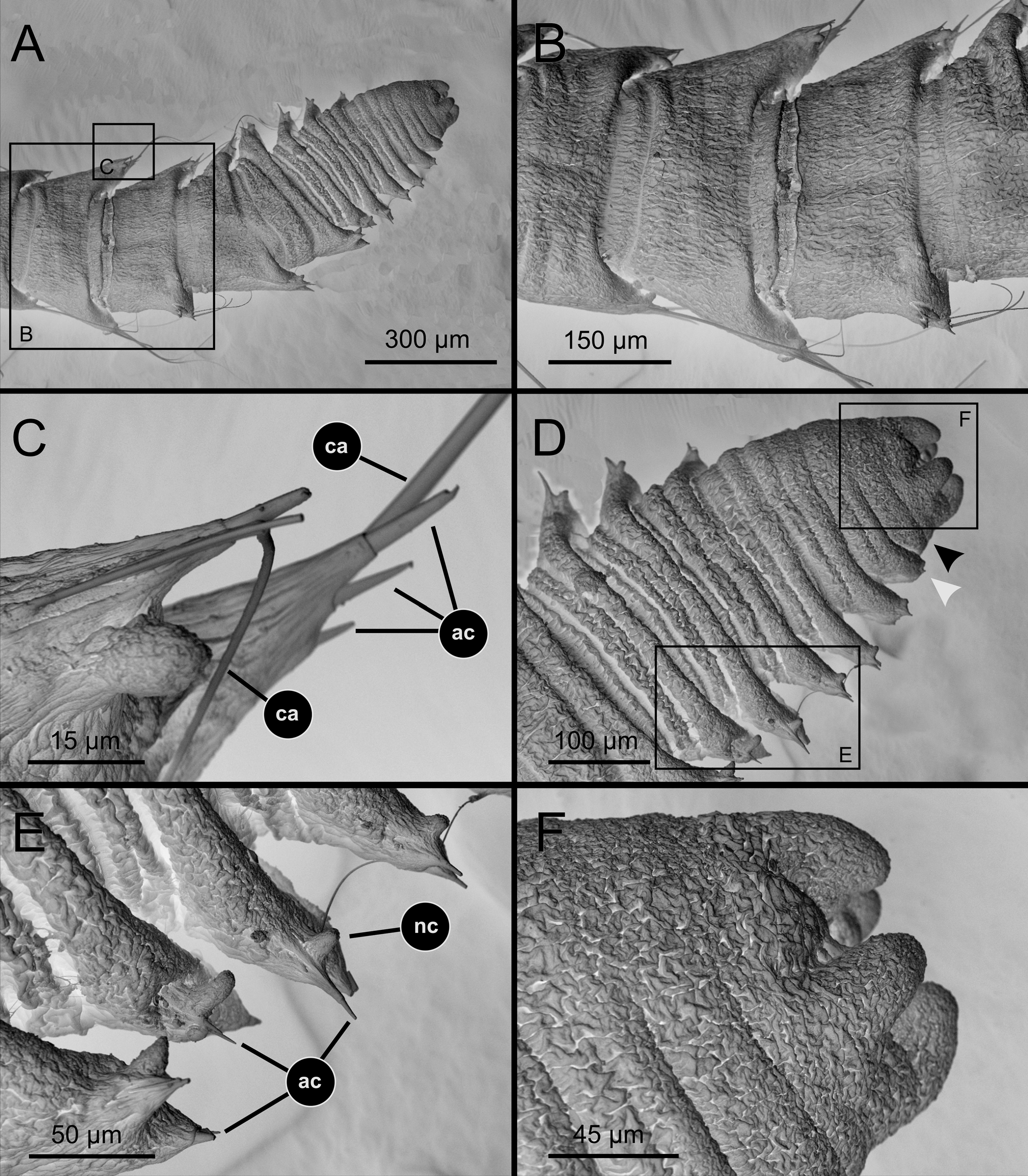
SEM micrographs showed lateral ciliary spots at both sides of prostomium ( Fig. 4 View FIGURE 4 E–F), which might represent nuchal organs (see Discussion). Annulation of segments between parapodial rami follows this pattern: one ring between CH 1 and CH 2, two rings between CH 2– CH 6(5) and three rings from CH 6(5) ( Figs 4 View FIGURE 4 A–D, 5A–B); annulation is less defined in pre-pygidial region ( Fig. 6 View FIGURE 6 D). Annulation is lost when segments are conspicuously elongated ( Fig. 6 View FIGURE 6 A–B). Notopodial papilla is always present from CH 1 ( Fig. 5 View FIGURE 5 C). Neuropodial papilla was not observed. Capillary and acicular chaetae are present in both rami from CH 1 ( Fig. 5 View FIGURE 5 C–F). Capillaries are long and numerous (9–10 per bundle) in the first 5–6 chaetigers ( Fig. 4 View FIGURE 4 A, C); acicular chaetae numbering up to 3 per rami.
Last 5–6 posterior segments suddenly become much shorter acquiring a crowded appearance ( Fig. 6 View FIGURE 6 A, D); these look achaetous but show small lateral projections (= small parapodia; Fig. 6 View FIGURE 6 D, black arrowhead). In some cases, body shows an inflated region just before the pygidium (see Blake 1985) which is probably a fixation artefact ( Fig. 2 View FIGURE 2 F). The pygidium is provided with four lobes ( Figs 3 View FIGURE 3 D–E, 6D, F).
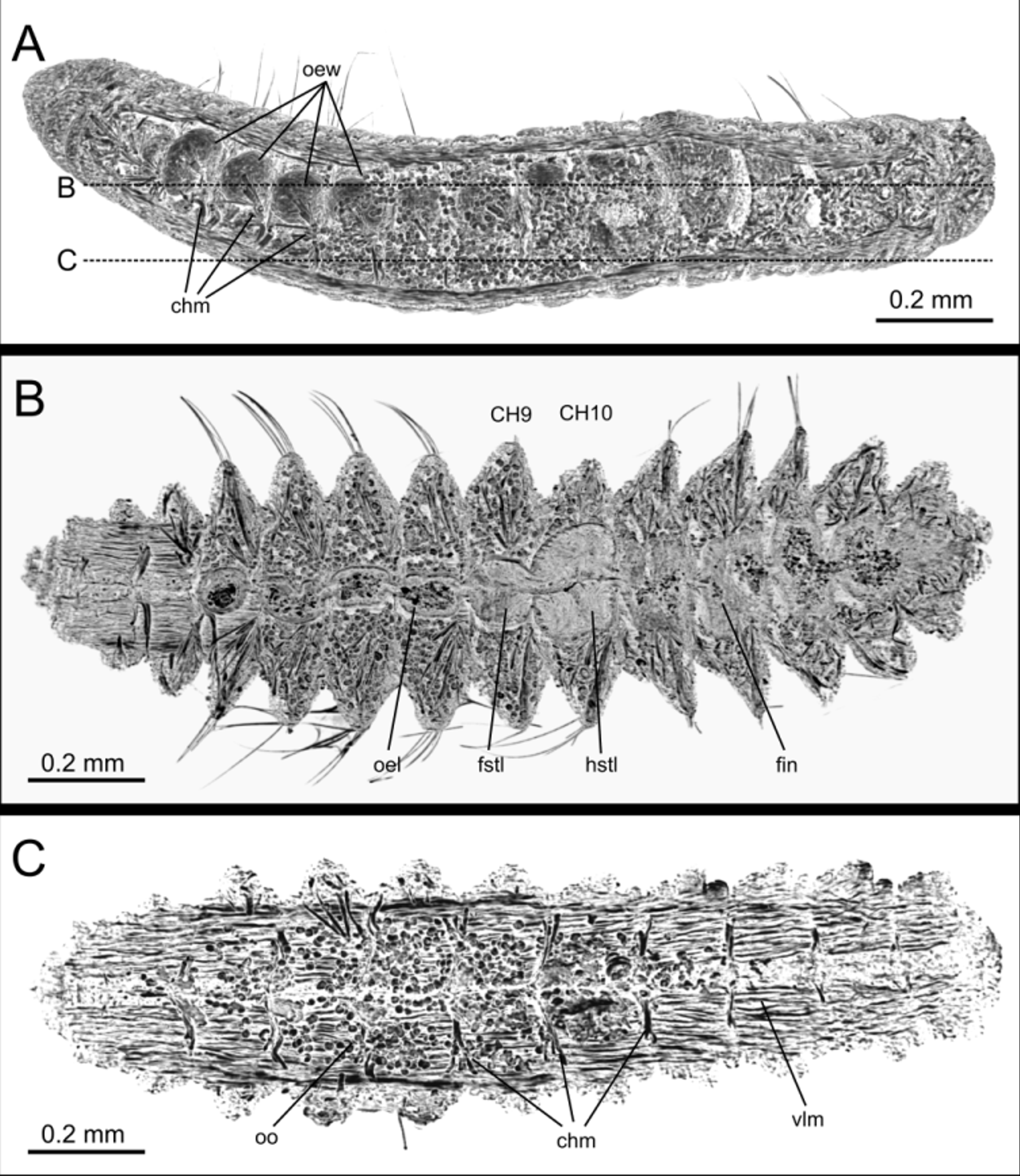
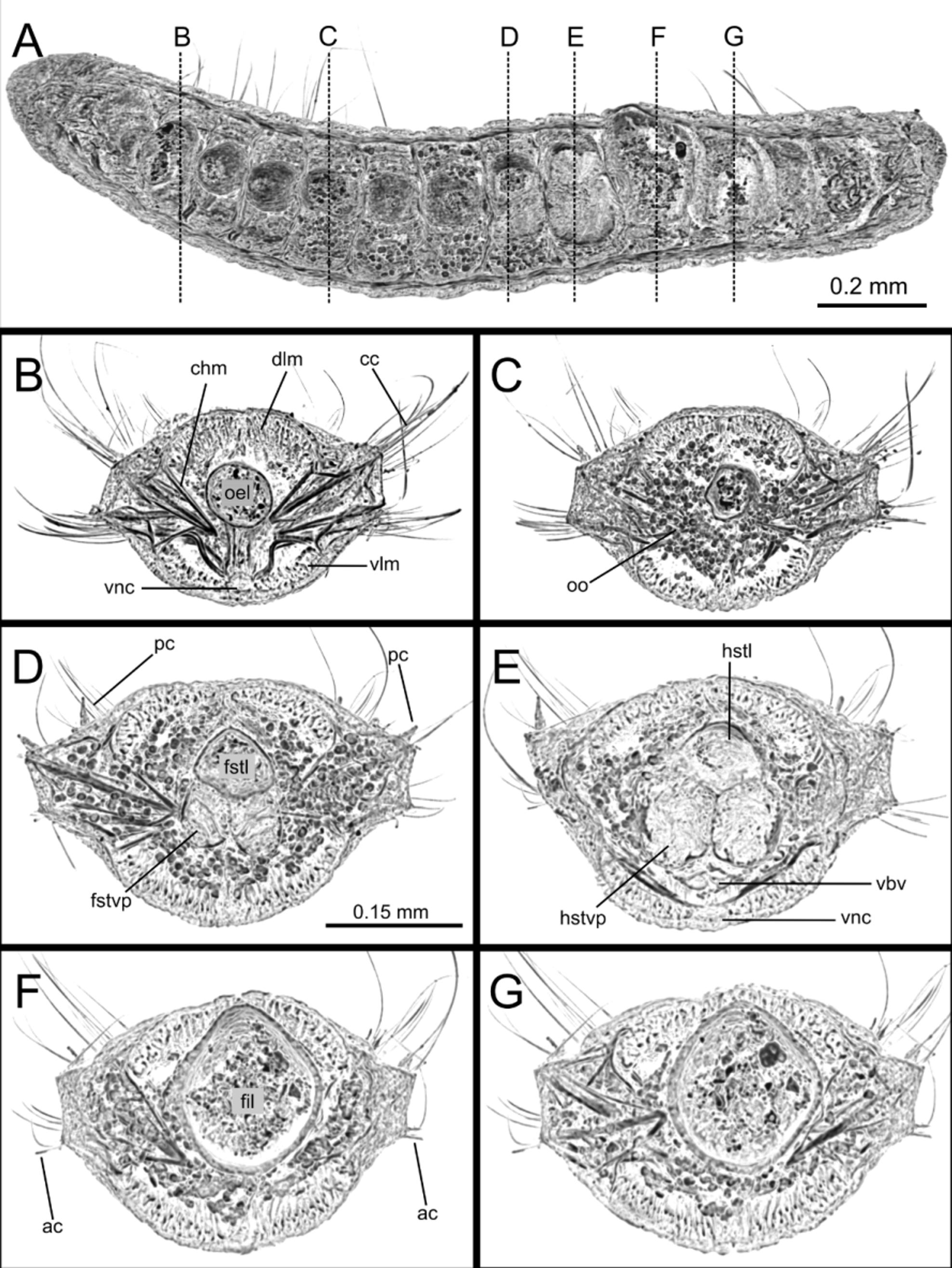
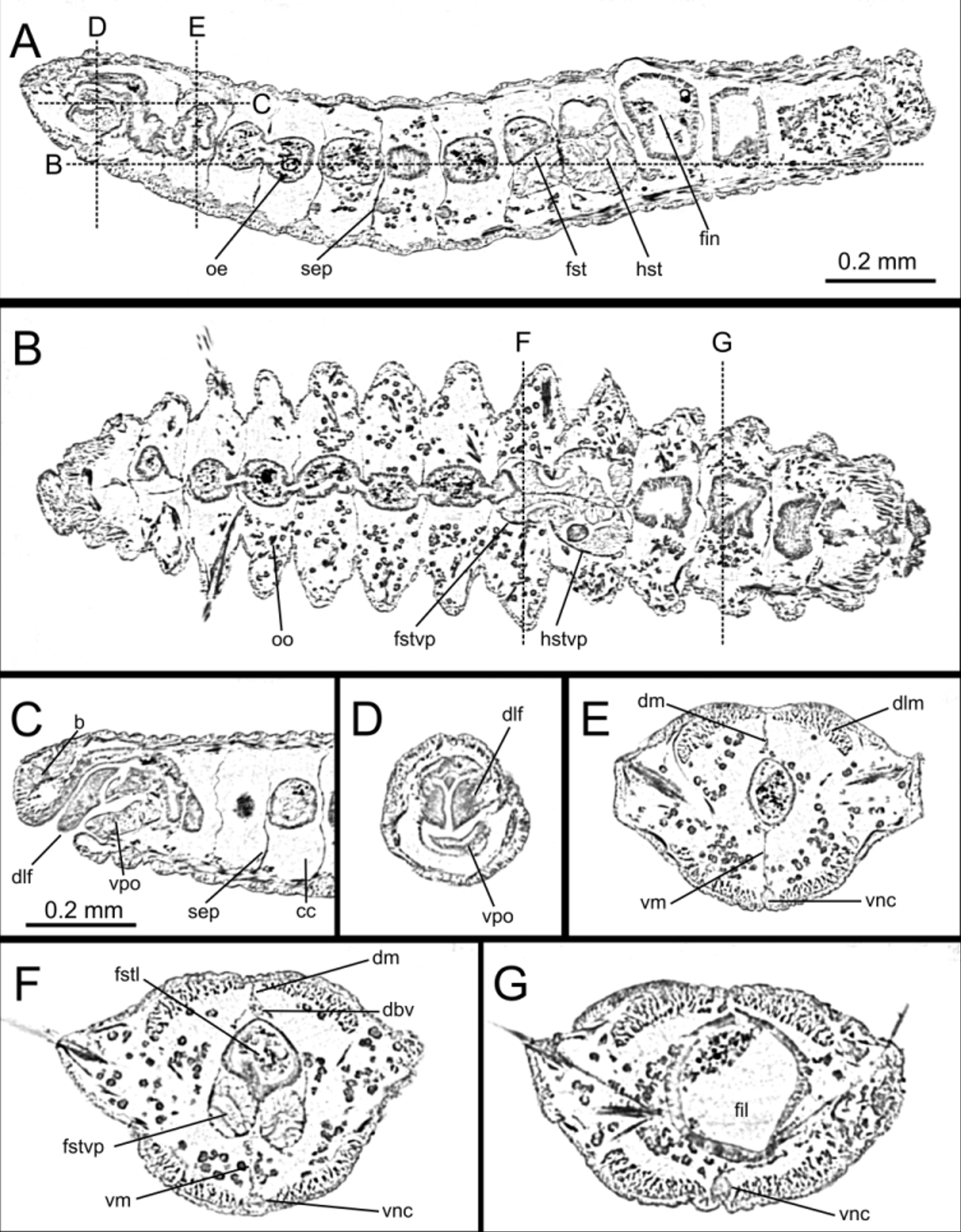
Gross internal anatomy. The study of three paratypes (IINH 35673) under the µCT allowed exploring for the first time the gross internal anatomy of any species of the genus Orbiniella ( Figs. 7–9 View FIGURE 7 View FIGURE 8 View FIGURE 9 ). The main findings were: (1) Body general cavity is well separated by septa ( Figs 7 View FIGURE 7 A, 8A, 9A, C), and is mostly filled by the digestive system ( Figs 7 View FIGURE 7 B, 8A, 9B) and maturing sexual cells (oocytes) ( Figs 7 View FIGURE 7 A–C, 8A, C–D, 9B, E–F). (2) Digestive system shows conspicuous regionalization ( Figs 7 View FIGURE 7 B, 9B) and is suspended by developed dorsal and ventral mesenteries ( Fig. 9 View FIGURE 9 E):
Species Eyes Nuchal Achaetous segments Segmental annulation Capillary chaetae Preanal Pygidium
organs segments2
. minuta Day, 1954 View in CoL no n.d. buccal segment + biannulated crenulated 0[2] one pair of rounded lobes
second segment
Extracted from the drawing because no explicit mention of this character is found in the original description. Data in brackets from Gillet (1999).
“unsegmented pre-pygidial region”.
a. Anterior part (oesophagus) long and narrow, running approximately from anterior end to CH 8 ( Figs 8 View FIGURE 8 A–B, 9E). Buccal cavity with two dorso-lateral folds and ventral pharyngeal organ.
b. Middle part (stomach) at level of CH 9 and CH 10 ( Figs 8 View FIGURE 8 D–E, 9F). Stomach trichambered, with dorsal space as prolongation of oesophagus and two defined ventral pouches (lumen not filled with sediment but apparently filled with sort of network); the latter seems to be connected to the stomach lumen at level of hind stomach.
c. Posterior part (intestine) with walls much thicker than those of stomach, running from CH 11 to end of body ( Figs 8 View FIGURE 8 F–G, 9G).
Most of the digestive tract lumen is filled with sediment including foraminiferans ( Fig. 8 View FIGURE 8 G). (3) Longitudinal body musculature well developed, organised in two dorsal and two ventral apparent bands ( Figs View FIGURE 7
7C, 8A–G, 9E–G), also including well defined musculature associated to parapodia and chaetae ( Figs 7 View FIGURE 7 A, C,
8B–D).
Other elements less discernible include dorsal and ventral blood vessels ( Figs 8 View FIGURE 8 E, 9E–G) and ventral nerve cord ( Figs 8 View FIGURE 8 B, E, 9E–F).
Etymology. The species is named after Danish polychaetologist Dr. Mary E. Petersen, who has recently passed away. Mary first suggested that these specimens might constitute a new species and her notes and drawings inspired this paper.
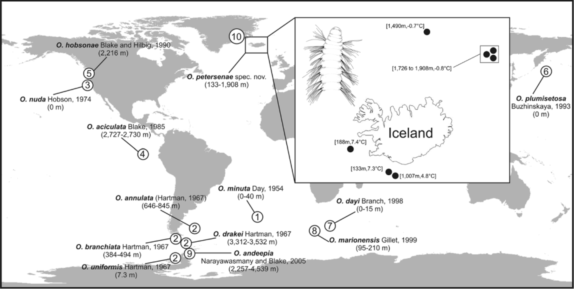
Distribution. Specimens were collected in two areas characterized by different predominant water masses ( Fig. 1 View FIGURE 1 ). Some of them were found on the shelf and slope (133 to 1,007 m depth) of SW Iceland, off Faxafloi Bay to South of Vestmannaeyjar Island, in warmer waters (4.8 to 7.4ºC). Most specimens were collected on the slope off NE Iceland, in Arctic colder, deeper waters (-0.7 to -0.8ºC; 1,490 to 1,915 m).
| MNCN |
Museo Nacional de Ciencias Naturales |
No known copyright restrictions apply. See Agosti, D., Egloff, W., 2009. Taxonomic information exchange and copyright: the Plazi approach. BMC Research Notes 2009, 2:53 for further explanation.
|
Kingdom |
|
|
Phylum |
|
|
Class |
|
|
Family |
|
|
Genus |
Orbiniella petersenae
| Parapar, Julio, Moreira, Juan & Helgason, Gudmundur Vidir 2015 |
O. andeepia
| Narayanaswamy and Blake 2005 |
O. marionensis
| Gillet 1999 |
O. dayi
| Branch 1998 |
O. plumisetosa
| Buzhinskaja 1993 |
O. hobsonae
| Blake and Hilbig 1990 |
O. aciculata
| Blake 1985 |
O. nuda
| Hobson 1974 |
O. annulata
| Hartman 1967 |
O. uniformis
| Hartman 1967 |
minuta
| Day 1954 |