
Paramblynotus anjiensis, Dong & Liu & Wang & Chen, 2018
|
publication ID |
https://doi.org/ 10.11646/zootaxa.4486.4.6 |
|
publication LSID |
lsid:zoobank.org:pub:A3DCA5C8-445A-45EB-8568-1B0B64E36917 |
|
DOI |
https://doi.org/10.5281/zenodo.5970029 |
|
persistent identifier |
https://treatment.plazi.org/id/7A2487FB-C464-FFAD-C1E5-FC10FB2BD6B3 |
|
treatment provided by |
Plazi |
|
scientific name |
Paramblynotus anjiensis |
| status |
sp. nov. |
Paramblynotus anjiensis , new species
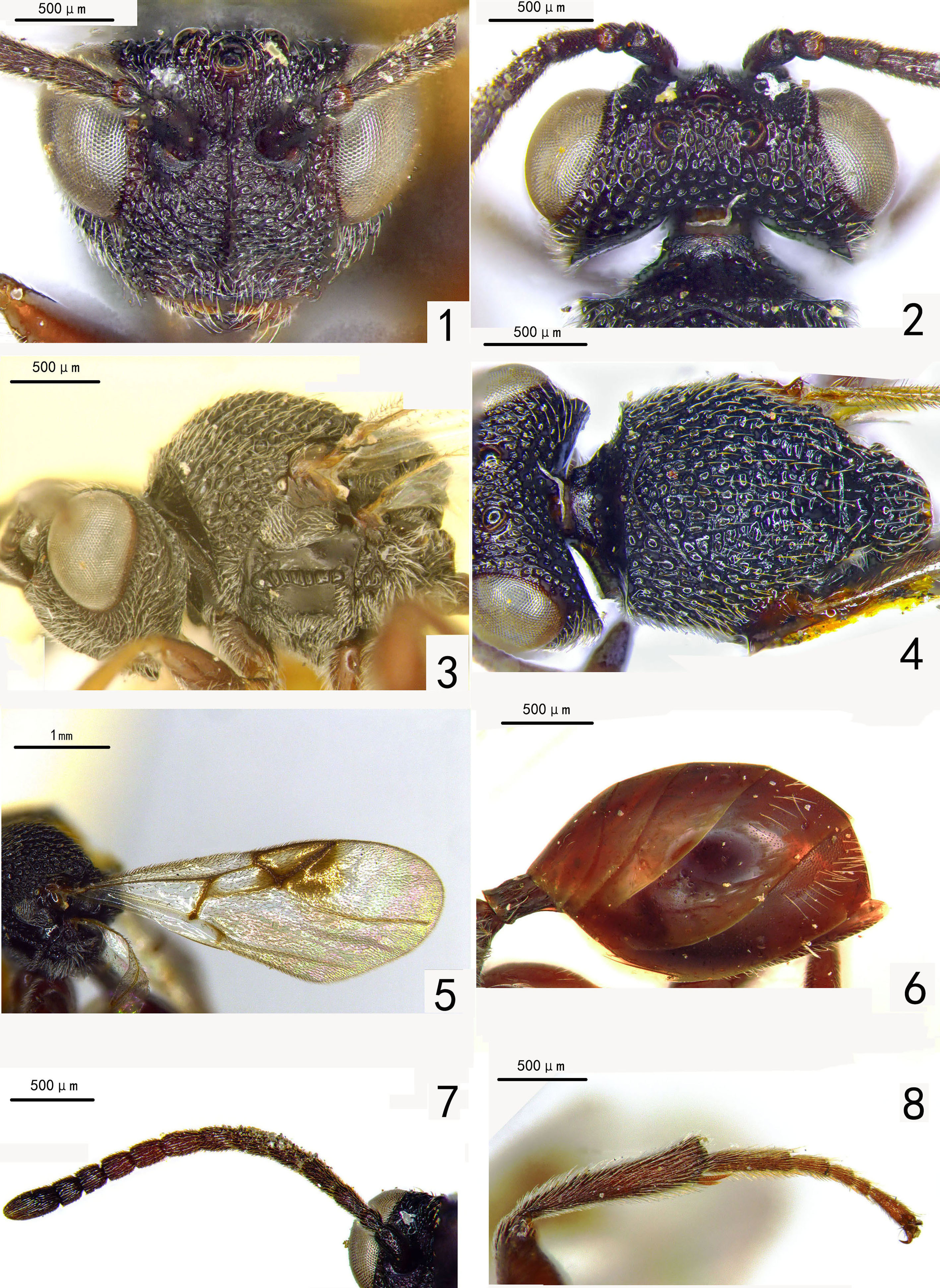
Figs 1–8 View FIGURES 1–8
Type material. HOLOTYPE female: CHINA, Zhejiang, Anji, Longwang Mt., Hong Wu , 1995. VII.20. PARATYPE . 1 female: CHINA, Zhejiang, Qingliangfeng, 2012.VIII (Rui Guo). (The type specimens are deposited in the Insect Collection of Zhejiang A & F University)
Etymology. This new species is named after the collection location of the holotype: Anji, Zhejiang Province of eastern China.
Diagnosis. Paramblynotus anjiensis is similar to P. hainanensis , but differs from the latter in: 1) median frontal carina is continuous from anterior ocellus to epistomal sulcus, distinctly raised, but not into a prominent lamella, above antennal sockets; 2) mesoscutellum posteriorly is not raised and not projected into two triangular processes.
Description. FEMALE (holotype). Body length 3.5–4.5 mm. Antenna 13-segmented, flagellum filiform. Fore wing ( Fig. 5 View FIGURES 1–8 ) with Rs+M arising from middle of basal vein. Marginal cell 3.0 times as long as wide and nearly as long as submarginal cell.
Coloration. Head and antenna dark brown. Mesosoma black. Legs and metasoma yellow-brown. Fore wing light gray,tinted yellow to yellowish brown along margin and veins, with a deep ferruginous macula covering marginal cell and part of third cubital cell behind marginal cell.
Head ( Figs. 1–2 View FIGURES 1–8 ). Vertex foveate-reticulate. Eye prominent, distinctly extended laterally beyond outer margin of gena. Ocellar plate slightly raised, foveate-reticulate, defined laterally by weak carina. Median frontal carina continuous from anterior ocellus to epistomal sulcus, distinctly raised, but not into a prominent lamella, above antennal sockets. Dorsal frons foveate-reticulate; antennal scrobe deeply depressed, densely punctate, foveate posteriorly, well defined laterally by carina. Gena foveate-reticulate and punctate, sparsely pubescent, horizontally costate posteriorly. Ventral frons punctate, foveate-reticulate with distinct transverse rugosity; clypeus densely punctate and longitudinally carinate; clypeo-pleurostomal sulcus and epistomal sulcus forming smoothly curved arch. Anterior tentorial pits small. Lateral occipital carina not reaching posterior part of vertex. Occiput mostly shining smooth and foveate above, with long setae laterally.
Mesosoma ( Figs. 3–4 View FIGURES 1–8 ). Anterior flange of pronotum finely transversely striate; submedian pronotal depressions separated medially. Anterior plate of pronotum glabrous, densely punctate dorsomedially. Pronotum dorsomedially slightly raised, but distinctly lower than mesoscutum; pronotal crest not raised medially. Lateral pronotal carina distinct, reaching pronotal crest dorsomedially. Lateral surface of pronotum foveate-reticulate and densely punctate. Dorsal pronotal area glabrate, present to middle of posterior margin of pronotum. Mesoscutum strongly arched dorsally, foveate-reticulate with foveae set in rows between interrupted transverse costae. Scutellar sulcus divided by median longitudinal carina. Mesoscutellum foveate-reticulate, posteriorly not raised or projected into two triangular processes. Axillar area without distinct pubescence. Mesopleural triangle pubescent, well defined ventrally by smoothly curved carina. Median mesopleural impression percurrent, transversely costate medially; upper mesopleuron glabrous except finely punctate anteriorly; lower mesopleuron glabrous, conspicuously pubescent ventrally. Metepisternum areolate-reticulate in dorsal part and conspicuously pubescent and densely punctate ventrally. Lateral propodeal carina abruptly curved medially; median propodeal area areolatereticulate; median longitudinal carina percurrent, crossed by submedian transverse carina.
Petiole 2.5 times as long as wide in lateral view. Tergum 8 not exposed; relative length of T3–7: 2.1:1.0:1.3:2.8:2.2; T3–5 glabrous; T6 and T7 finely punctate, each with a middle row of pubescence dorsolaterally. Apical teeth of metatibia long, slender, pointed apically. Length of 1 st metatarsomere 0.55 times combined length of 2–5mt.
MALE. Unknown.
Distribution. China: Zhejiang (Oriental region).
Comments. The species appears to be much smaller than P. hainanensis , the species it resembles most. However, more specimens are needed to conclude whether the difference is statistically meaningful.
No known copyright restrictions apply. See Agosti, D., Egloff, W., 2009. Taxonomic information exchange and copyright: the Plazi approach. BMC Research Notes 2009, 2:53 for further explanation.