
Priceiella (Thescelovora) fuscicaena Gustafsson, Clayton
|
publication ID |
https://doi.org/ 10.11646/zootaxa.4382.3.1 |
|
publication LSID |
lsid:zoobank.org:pub:4BE1AB50-46E7-402D-9E72-A45D78352E2B |
|
DOI |
https://doi.org/10.5281/zenodo.5995563 |
|
persistent identifier |
https://treatment.plazi.org/id/E11BB55A-B745-FFC3-FF76-908CFAE06DFA |
|
treatment provided by |
Plazi |
|
scientific name |
Priceiella (Thescelovora) fuscicaena Gustafsson, Clayton |
| status |
|
Priceiella (Thescelovora) fuscicaena Gustafsson, Clayton , & Bush, new species
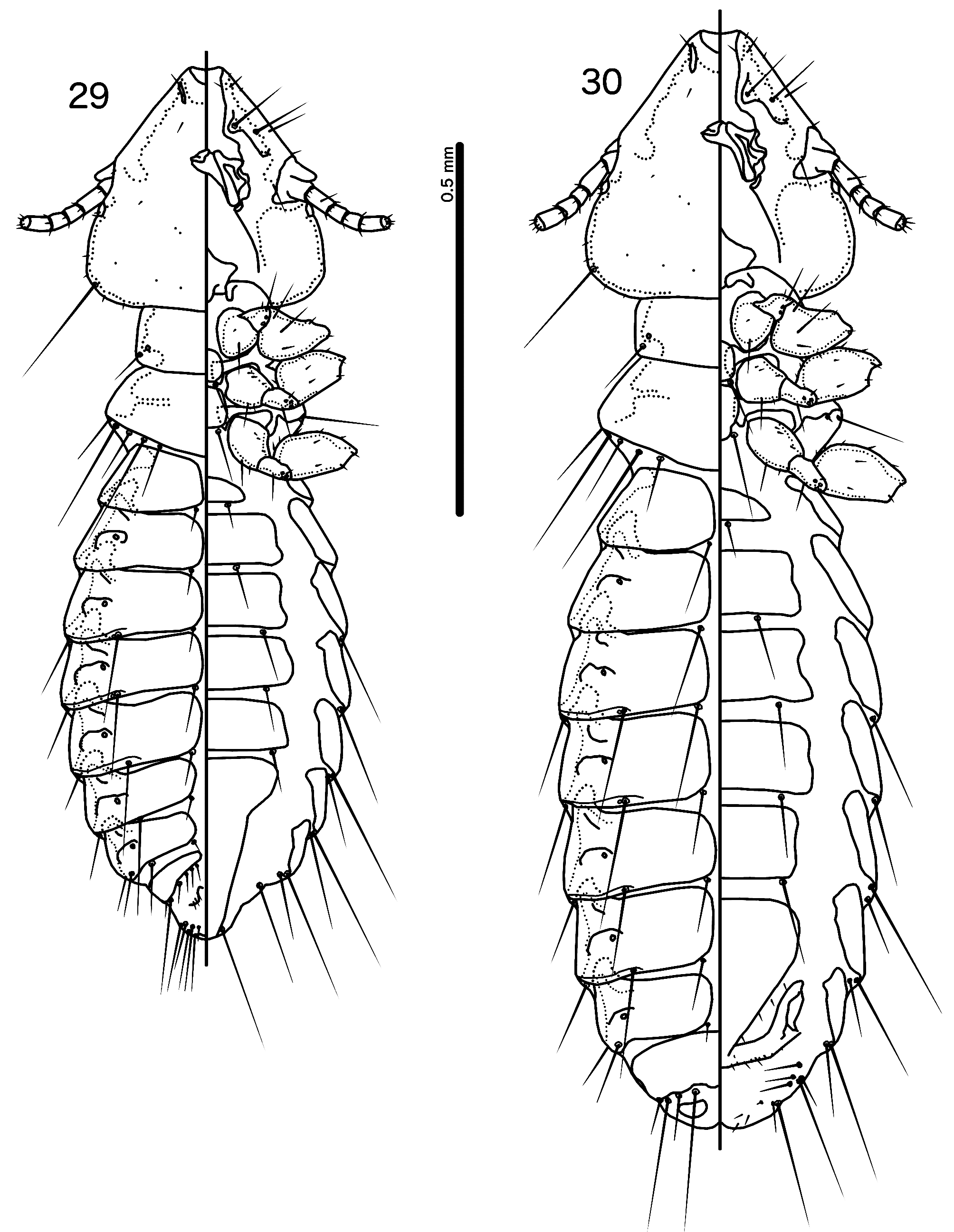
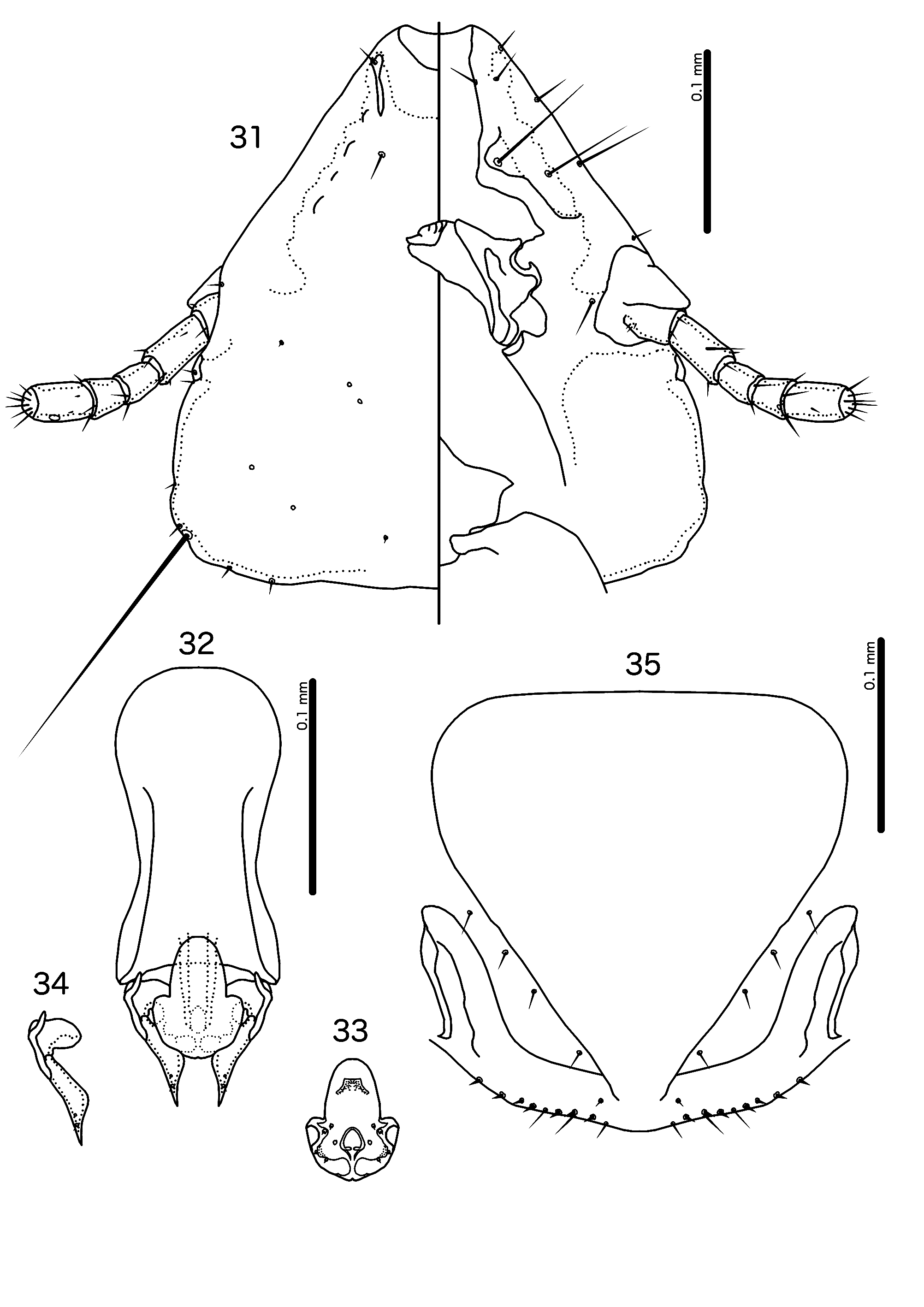
( Figs 29–35 View FIGURES 29–30 View FIGURES31–35 )
Type host. Malacopteron magnum magnum Eyton, 1839—rufous-crowned babbler ( Pellorneidae ).
Type locality. Terengganu, elev. 140 ft., 102° 40’E. 5° 28’ N, Malaysia GoogleMaps .
Other hosts. Malacopteron cinereum cinereum Eyton, 1839 —scaly-crowned babbler ( Pellorneidae ).
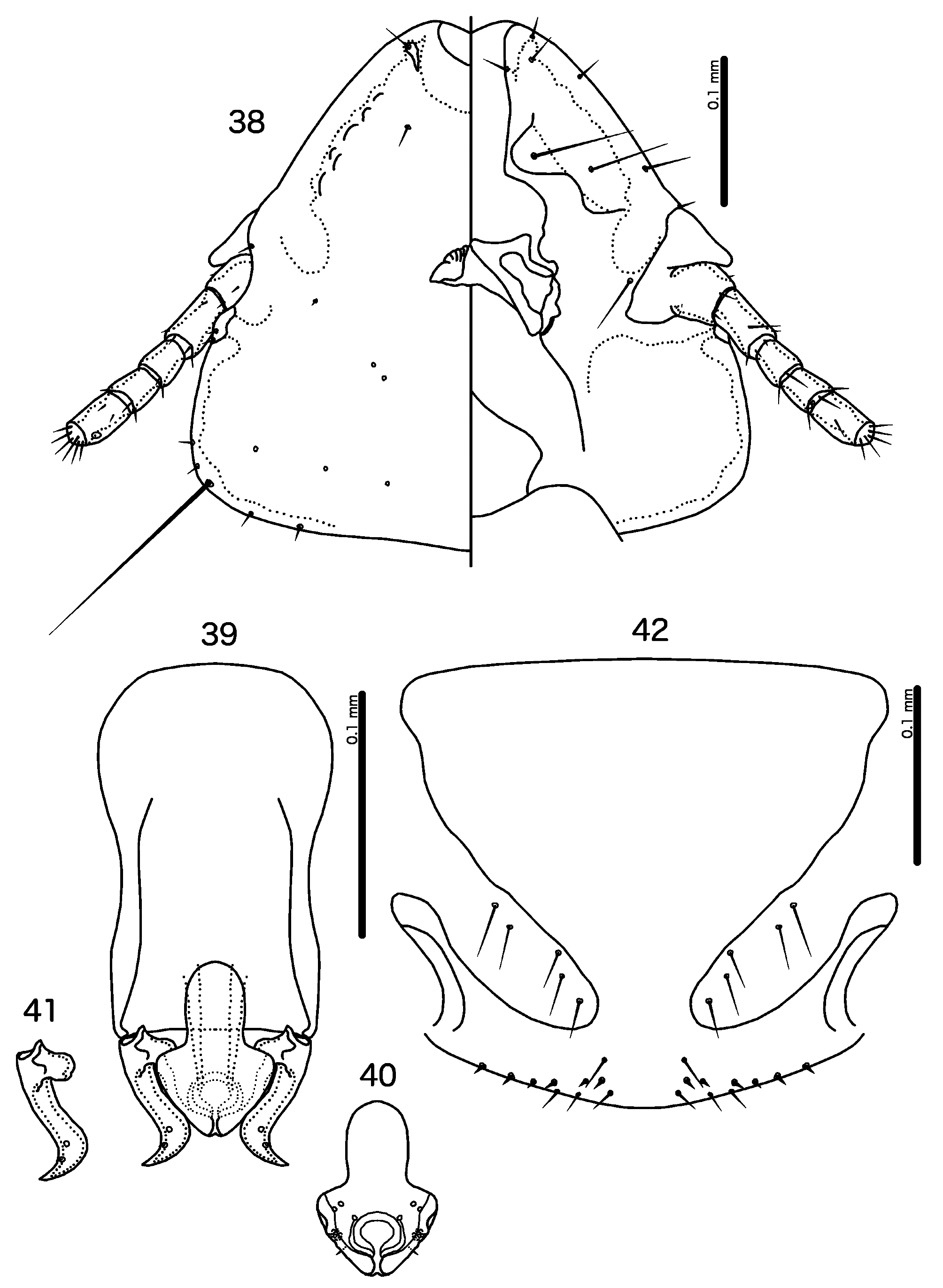
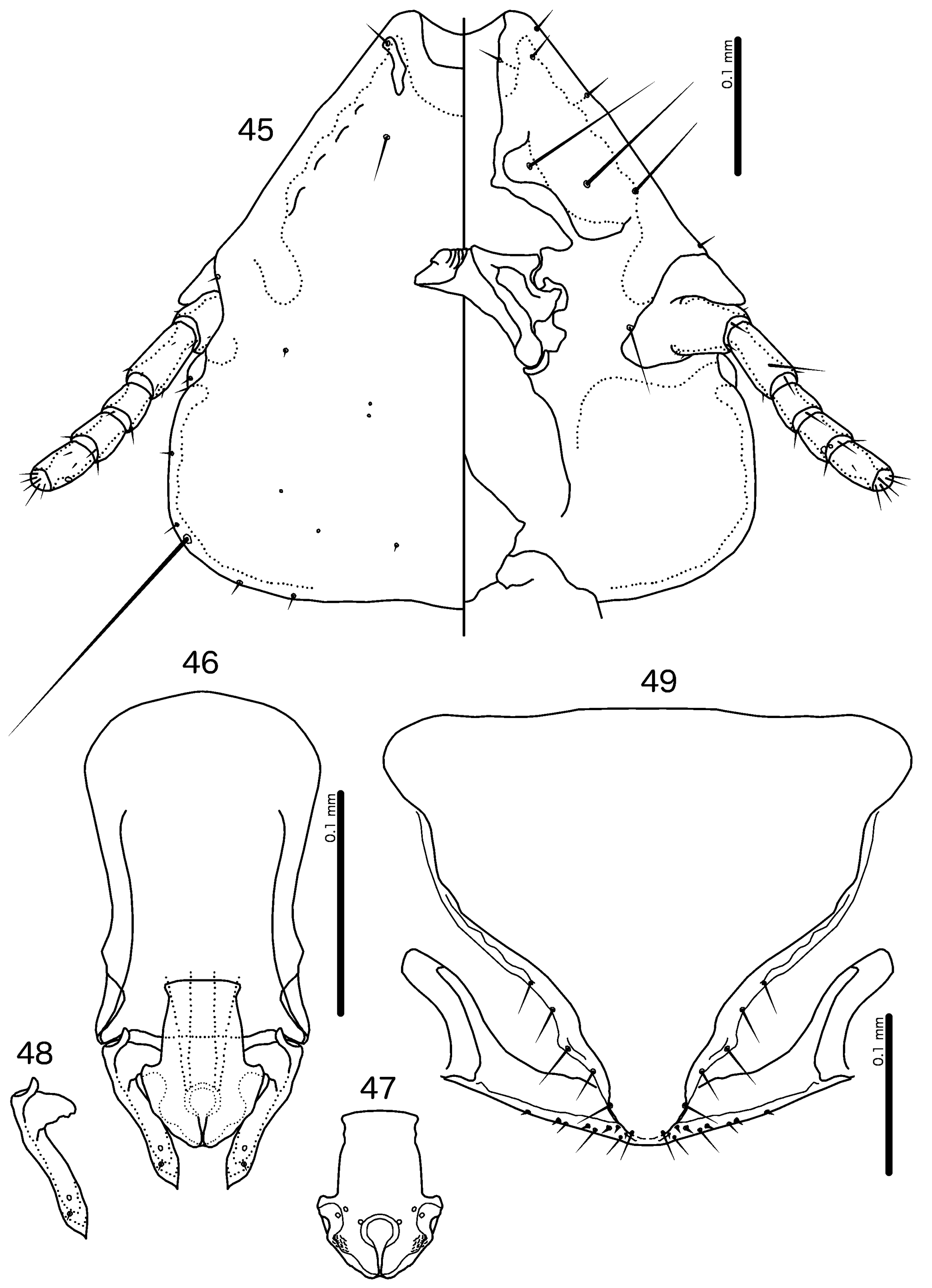
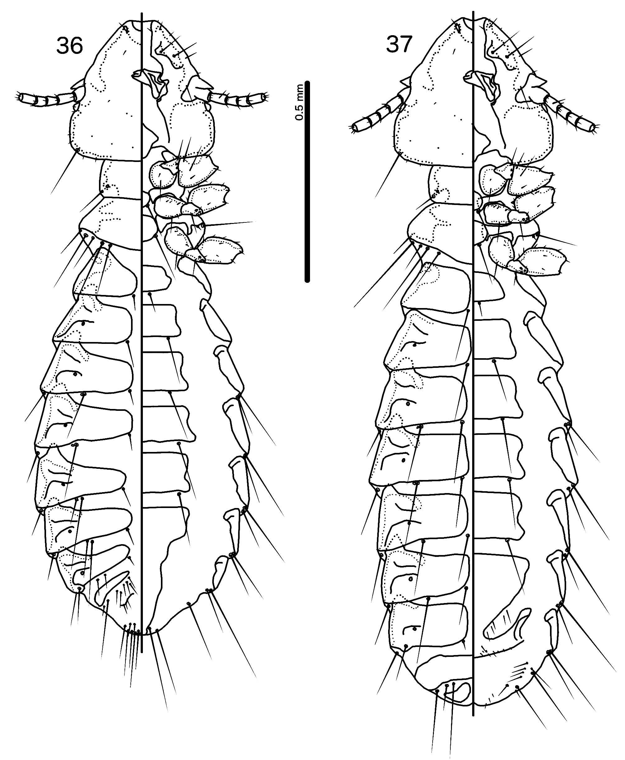
Diagnosis. The general structure of the male genitalia places P. (T.) fuscicaena n. sp. ( Figs 32–34 View FIGURES31–35 ) close to P. (T.) coleyae n. sp. ( Figs 39–41 View FIGURES 38–42 ). Both of these species have a rounded proximal mesosome and mesosomal lobes with broad marginal thickening ( Figs 33 View FIGURES31–35 , 40 View FIGURES 38–42 ), which separate both from the angular proximal mesosome and mesosomal lobes with slender marginal thickening of P. (T.) macrocephala n. sp. ( Fig. 47 View FIGURES 45–49 ). However, the elongated head shape and extended dorsal preantennal suture ( Figs 31 View FIGURES31–35 , 45 View FIGURES 45–49 ) and absence of aps on male tergopleurites VI–VII ( Figs 29 View FIGURES 29–30 , 43 View FIGURES 43–44 ) places P. (T.) fuscicaena closer to P. (T.) macrocephala than to P. (T.) coleyae , which has a shorter head and less extensive dorsal preantennal suture ( Fig. 38 View FIGURES 38–42 ) and aps on male tergopleurites VI–VII ( Fig. 36 View FIGURES 36–37 ). It is not clear which of these species is the closest relative to P. (T.) fuscicaena .
Priceiella (Thescelovora) fuscicaena can be separated from P. (T.) coleyae on the following characters: preantennal head short and rounded in P. (T.) coleyae ( Fig. 38 View FIGURES 38–42 ) but more elongated in P. (T.) fuscicaena ( Fig. 31 View FIGURES31–35 ); dorsal preantennal suture extending at least half-way between dsms and ads in P. (T.) fuscicaena ( Fig. 31 View FIGURES31–35 ) but less than half-way between these setae in P. (T.) coleyae ( Fig. 38 View FIGURES 38–42 ); aps absent on male tergopleurites VI–VII in P. (T.) fuscicaena ( Fig. 29 View FIGURES 29–30 ) but present there in P. (T.) coleyae ( Fig. 36 View FIGURES 36–37 ); basal apodeme slender, notably constricted at mid-length in P. (T.) fuscicaena ( Fig. 32 View FIGURES31–35 ) but broader and less or not constricted in P. (T.) coleyae ( Fig. 39 View FIGURES 38–42 ); distal mesosome roughly rounded in P. (T.) fuscicaena ( Fig. 33 View FIGURES31–35 ) but convergent to medial point in P. (T.) coleyae ( Fig. 40 View FIGURES 38–42 ); proximal mesosome with ventral rugose area in P. (T.) fuscicaena ( Fig. 33 View FIGURES31–35 ) but without such area in P. (T.) coleyae ( Fig. 40 View FIGURES 38–42 ); parameres parallel distally in P. (T.) fuscicaena ( Fig. 34 View FIGURES31–35 ) but divergent distally in P. (T.) coleyae ( Fig. 41 View FIGURES 38–42 ); vos longer in P. (T.) coleyae ( Fig. 42 View FIGURES 38–42 ) than in P. (T.) fuscicaena ( Fig. 35 View FIGURES31–35 ) but vulval chaetotaxy otherwise similar.
Priceiella (Thescelovora) fuscicaena can be separated from P. (T.) macrocephala on the following characters: size (see measurements and cf. Figs 29–30 View FIGURES 29–30 , 43–44 View FIGURES 43–44 ); both sexes of P. (T.) macrocephala with 2 sts on abdominal segment VI ( Figs 43–44 View FIGURES 43–44 ), but only 1 sts on abdominal segment VI in both sexes of P. (T.) fuscicaena ( Figs 29–30 View FIGURES 29–30 ); male with 1 tps on tergopleurite VIII in P. (T.) fuscicaena ( Fig. 29 View FIGURES 29–30 ) but 2 tps in P. (T.) macrocephala ( Fig. 43 View FIGURES 43–44 ); basal apodeme slender, notably constricted at mid-length in P. (T.) fuscicaena ( Fig. 32 View FIGURES31–35 ) but broader and less or not constricted in P. (T.) macrocephala ( Fig. 46 View FIGURES 45–49 ); proximal mesosome rounded in P. (T.) fuscicaena ( Fig. 33 View FIGURES31–35 ) but rectangular with roughly flattened anterior margin in P. (T.) macrocephala ( Fig. 47 View FIGURES 45–49 ); proximal mesosome with ventral rugose area in P. (T.) fuscicaena ( Fig. 33 View FIGURES31–35 ) but without such area in P. (T.) macrocephala ( Fig. 47 View FIGURES 45–49 ); marginal thickening of mesosomal lobes broad distally in P. (T.) fuscicaena ( Fig. 33 View FIGURES31–35 ) but slender distally in P. (T.) macrocephala ( Fig. 47 View FIGURES 45–49 ); vos much longer in P. (T.) macrocephala ( Fig. 49 View FIGURES 45–49 ) than in P. (T.) fuscicaena ( Fig. 35 View FIGURES31–35 ), but vulval chaetotaxy otherwise similar.
Description. Both sexes. Head pentagonal ( Fig. 31 View FIGURES31–35 ). Frons flat to shallowly concave. Lateral margins of preantennal head straight to slightly concave. Dorsal preantennal suture reaches dsms but only half-way to ads. Head chaetotaxy as in Fig. 31 View FIGURES31–35 . Coni reach distal margin of scape. Pteronotum with 5 mms on each side ( Figs 29– 30 View FIGURES 29–30 ). Pigmentation pale and most of body near translucent; marginal carina, head nodi, gular plate, proepimera, metepisterna and pleural incrassations pale brown; sternal plate VI and subgenital plate very pale brown.
Male. Abdominal plates and chaetotaxy as in Fig. 29 View FIGURES 29–30 ; aps absent on tergopleurites VI–VII. Male genitalia as in Figs 32–34 View FIGURES31–35 . Basal apodeme rounded, slender, constricted at mid-length ( Fig. 32 View FIGURES31–35 ). Proximal mesosome half-oval, narrow, with rugose ventral area ( Fig. 33 View FIGURES31–35 ). Mesosomal lobes gently rounded; distal end of mesosome rounded. Ventral nodi slightly rugose submarginally. Gonopore open only distally; 2 ames sensilla on each side near anterolateral corners of mesosomal lobes; 1 pmes sensillus on each side of posterior end of gonopore; 2 pmes sensilla on each side distal to gonopore, 1 lateral to rugose nodi and 1 medio-posterior to rugose nodi. Parameral heads large, rounded medially ( Fig. 34 View FIGURES31–35 ). Parameral blades slender, convergent. Measurements ex Malacopteron magnum magnum (n = 4, except TL where n = 3): TL = 1.15–1.42; HL = 0.32–0.34; HW = 0.32–0.35; PRW = 0.20–0.21; PTW = 0.27–0.30; AW = 0.37–0.44. Measurements ex Malacopteron cinereum cinereum (n = 1): TL = 1.24; HL = 0.33; HW = 0.33; PRW = 0.19; PTW = 0.28; AW = 0.39.
Female. Abdominal plates and chaetotaxy as in Fig. 30 View FIGURES 29–30 . Vulval margin gently rounded ( Fig. 35 View FIGURES31–35 ), with 2–3 slender vms and 6–7 thorn-like vss on each side; 4–7 short, slender vos on each side; distal vos much shorter than proximal vos and located just anterior to vss. Measurements ex Malacopteron magnum magnum (n = 3): TL = 1.43–1.47; HL = 0.34–0.35; HW = 0.35–0.36; PRW = 0.21; PTW = 0.31; AW = 0.42–0.47. Measurements ex Malacopteron cinereum cinereum (n = 1): TL = 1.60; HL = 0.37; HW = 0.38; PRW = 0.22; PTW = 0.31; AW = 0.49.
Etymology. The species epithet is derived from Latin “ fuscus ” for “brown” and “ caeno ” for “to dine”, referring to the brownish color of both host species.
Type material. Ex Malacopteron magnum magnum: Holotype Ƌ, Terengganu, elev. 140 ft., 102° 40’E, 5° 28’ N, Malaysia, 26 Feb. 1974, Gn. Lawit Expedition, Brit. Mus. 1974–2 ( NHML) GoogleMaps . Paratypes: 2♂, 2♀, same data as holotype ( NHML) GoogleMaps .
Additional material examined (non-types). Ex Malacopteron magnum magnum: 1♂, 1♀ Subang, Malaysia, 7 Mar. 1962, M-00957 ( OSUS).
Ex Malacopteron cinereum cinereum : 1♂, 1♀ Gombak, Malaysia, 14 Feb. 1963, M-02390 ( OSUS).
Remarks. No significant differences have been found between the samples from the two host species.

No known copyright restrictions apply. See Agosti, D., Egloff, W., 2009. Taxonomic information exchange and copyright: the Plazi approach. BMC Research Notes 2009, 2:53 for further explanation.
|
Kingdom |
|
|
Phylum |
|
|
Class |
|
|
Order |
|
|
Family |
|
|
Genus |