
Priceiella (Thescelovora) macrocephala Gustafsson, Clayton
|
publication ID |
https://doi.org/ 10.11646/zootaxa.4382.3.1 |
|
publication LSID |
lsid:zoobank.org:pub:4BE1AB50-46E7-402D-9E72-A45D78352E2B |
|
DOI |
https://doi.org/10.5281/zenodo.5995567 |
|
persistent identifier |
https://treatment.plazi.org/id/E11BB55A-B75F-FFC4-FF76-9180FEF068E5 |
|
treatment provided by |
Plazi |
|
scientific name |
Priceiella (Thescelovora) macrocephala Gustafsson, Clayton |
| status |
|
Priceiella (Thescelovora) macrocephala Gustafsson, Clayton , & Bush, new species
( Figs 43–49 View FIGURES 43–44 View FIGURES 45–49 )
Type host. Megapomatorhinus hypoleucos wrayi Sharpe, 1887 —large scimitar-babbler ( Timaliidae ).
Type locality. Gunong Benom, elev. 6000 ft., Malaysia.
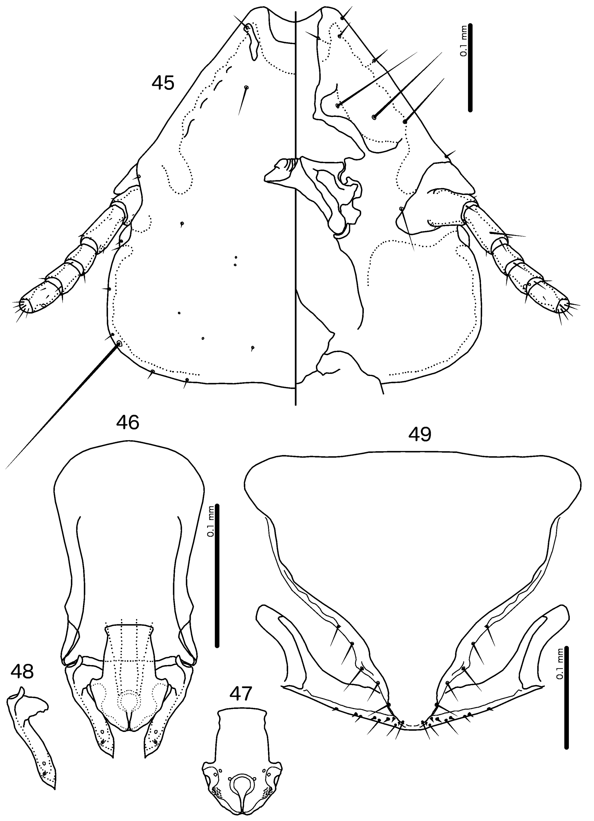
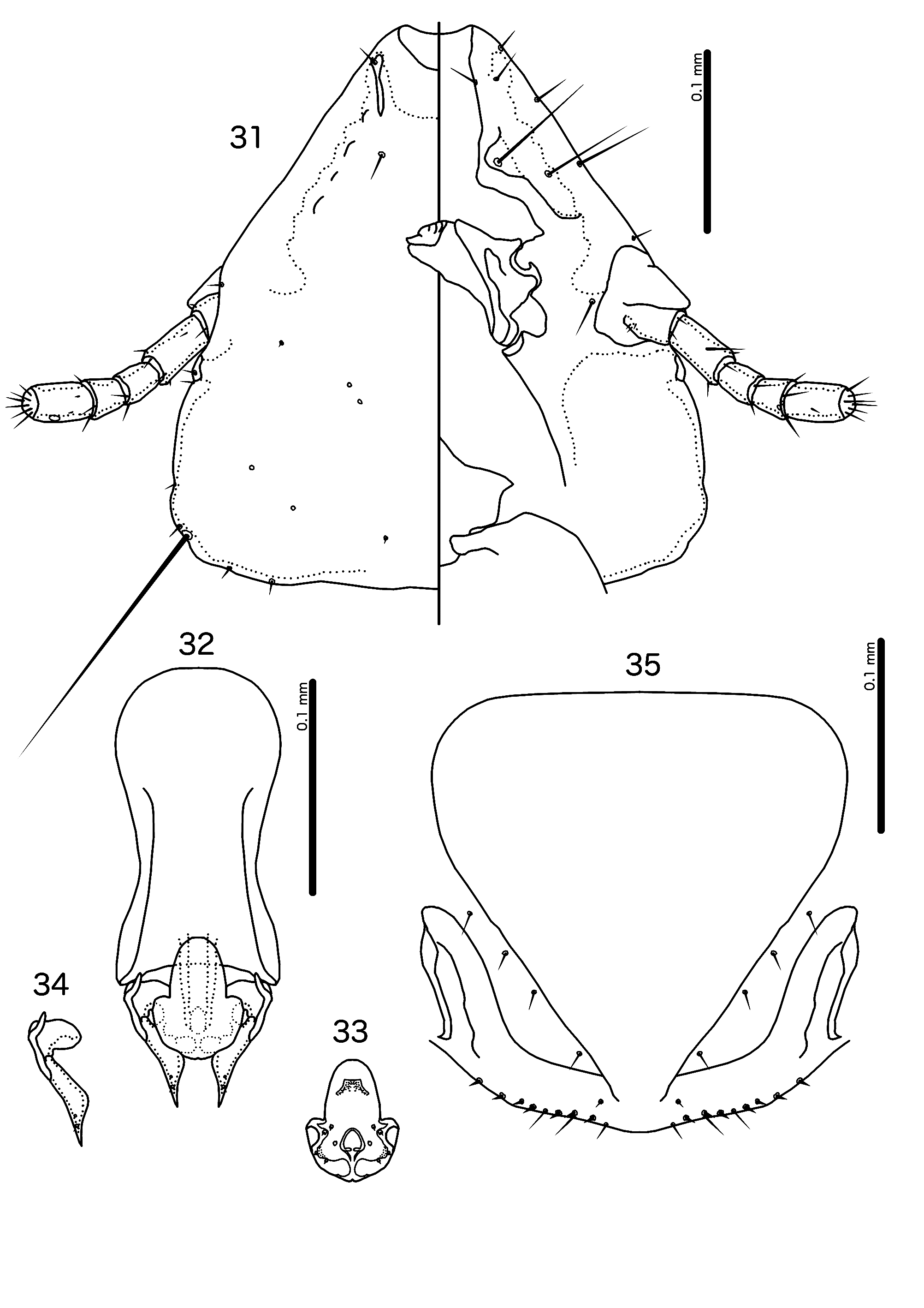
Diagnosis. Priceiella (Thescelovora) macrocephala n. sp. is most similar to P. (T.) fuscicaena n. sp., with which it shares the following characters: preantennal area long, with straight lateral margins ( Figs 31 View FIGURES31–35 , 45 View FIGURES 45–49 ); dorsal preantennal suture reaching at least half-way between dsms and ads ( Figs 31 View FIGURES31–35 , 45 View FIGURES 45–49 ); parameres not divergent distally ( Figs 34 View FIGURES31–35 , 48 View FIGURES 45–49 ); aps absent from male tergopleurites VI–VII. These two species can be separated on the following characters: proximal mesosome rectangular without rugose ventral area in P. (T.) macrocephala ( Fig. 47 View FIGURES 45–49 ), but rounded and with rugose ventral area in P. (T.) fuscicaena ( Fig. 33 View FIGURES31–35 ); marginal thickening of mesosomal lobes slender in P. (T.) macrocephala ( Fig. 47 View FIGURES 45–49 ), but broad in P. (T.) fuscicaena ( Fig. 33 View FIGURES31–35 ); pmes not visible near rugose nodi in P. (T.) macrocephala ( Fig. 47 View FIGURES 45–49 ), but visible in P. (T.) fuscicaena ( Fig. 33 View FIGURES31–35 ); parameres tapering gradually, with distal ends slender in P. (T.) fuscicaena ( Fig. 34 View FIGURES31–35 ), but tapering only in distal end and with roughly same width for most of length in P. (T.) macrocephala ( Fig. 48 View FIGURES 45–49 ); vos longer in P. (T.) macrocephala ( Fig. 49 View FIGURES 45–49 ) than in P. (T.) fuscicaena ( Fig. 35 View FIGURES31–35 ), but vulval chaetotaxy otherwise similar.
Description. Both sexes. Head pentagonal ( Fig. 45 View FIGURES 45–49 ). Frons deeply concave. Lateral margins of preantennal head roughly straight. Dorsal preantennal suture reaches dsms and at least half-way to ads. Head chaetotaxy as in Fig. 45 View FIGURES 45–49 . Coni reach distal margin of scape. Base pigmentation very pale brown; marginal and marginal temporal carinae, head nodi, flagellomeres I–II, proepimera, metepisterna and pleural incrassations dark reddish brown; mandibular framework, margins of antennal sockets, gular plate, flagellomere III and sternal and subgenital plates medium brown; sternal plates paler medianly.
Male. Pteronotum with 7–8 mms on each side ( Fig. 43 View FIGURES 43–44 ) (one male with 10 on one side). Abdominal plates and chaetotaxy as in Fig. 43 View FIGURES 43–44 ; aps absent on tergopleurites VI–VII. Male genitalia as in Figs 46–48 View FIGURES 45–49 . Basal apodeme broad, rectangular, slightly constricted at mid-length ( Fig. 46 View FIGURES 45–49 ). Proximal mesosome broad, with flat anterior margin ( Fig. 47 View FIGURES 45–49 ); typically widened proximally. Mesosomal lobes gently rounded to blunt medial point. Lateral thickening of mesosome sinuous. Ventral nodi rugose. Gonopore open only distally; 2 ames sensilla on each side near anterolateral corner of mesosomal lobes; 1 pmes sensilla on each side lateral to anterior end of gonopore. No lateral pmes discernable near rugose nodi, but these may be overlooked due to being sensilla. Parameral heads roughly triangular, with irregularly serrated posterior margin ( Fig. 48 View FIGURES 45–49 ). Parameral blades short, of roughly the same width for most of length; pst1–2 close together. Measurements ex Pomatorhinus hypoleucos wrayi (n = 6): TL = 1.62– 1.77; HL = 0.40–0.43; HW = 0.40–0.43; PRW = 0.24–0.26; PTW = 0.37–0.41; AW = 0.53–0.59.
Female. Pteronotum with 7 mms on each side ( Fig. 44 View FIGURES 43–44 ). Abdominal plates and chaetotaxy as in Fig. 44 View FIGURES 43–44 . Vulval margin gently rounded ( Fig. 49 View FIGURES 45–49 ) with 3–4 slender vms on each side (lateral vms much shorter than more medial vms), 5–6 thorn-like vss on each side; 5–7 slender vos on each side; distal vos medial to vss. Measurements ex Pomatorhinus hypoleucos wrayi (n = 6): TL = 1.83–2.01; HL = 0.42–0.44; HW = 0.43–0.46; PRW = 0.25–0.28; PTW = 0.39–0.42; AW = 0.57–0.65.
Etymology. The species epithet is derived from Greek “ makros ” for “large” and “ kefali ” for head, referring to its much larger head compared with closely related species.
Type material. Ex Pomatorhinus hypoleucos wrayi : Holotype Ƌ, Gunong Benom , elev. 6000 ft., Malaysia, 28 Mar. 1967, BA-40, Brit. Mus. 1967-400 ( NHML) . Paratypes: 5♂, 6♀, same data as holotype ( NHML) .
Additional material examined (non-types)
Ex Pomatorhinus hypoleucos wrayi : 1♂, 1♀, Mount Brinchang, Malaysia, 16 Mar. 1963, M-02691, 24716 on reverse ( OSUS).

No known copyright restrictions apply. See Agosti, D., Egloff, W., 2009. Taxonomic information exchange and copyright: the Plazi approach. BMC Research Notes 2009, 2:53 for further explanation.