
Sphaerodoropsis plurituberculata, Capa, María & Rouse, Greg W., 2015
|
publication ID |
https://doi.org/ 10.11646/zootaxa.4019.1.9 |
|
publication LSID |
lsid:zoobank.org:pub:D4ECFEE2-BF9D-4B5F-8128-EFAAC2B728C4 |
|
DOI |
https://doi.org/10.5281/zenodo.6096005 |
|
persistent identifier |
https://treatment.plazi.org/id/C22DFF62-FFF9-FF87-FF21-F9DDFB069FAC |
|
treatment provided by |
Plazi |
|
scientific name |
Sphaerodoropsis plurituberculata |
| status |
sp. nov. |
Sphaerodoropsis plurituberculata n. sp.
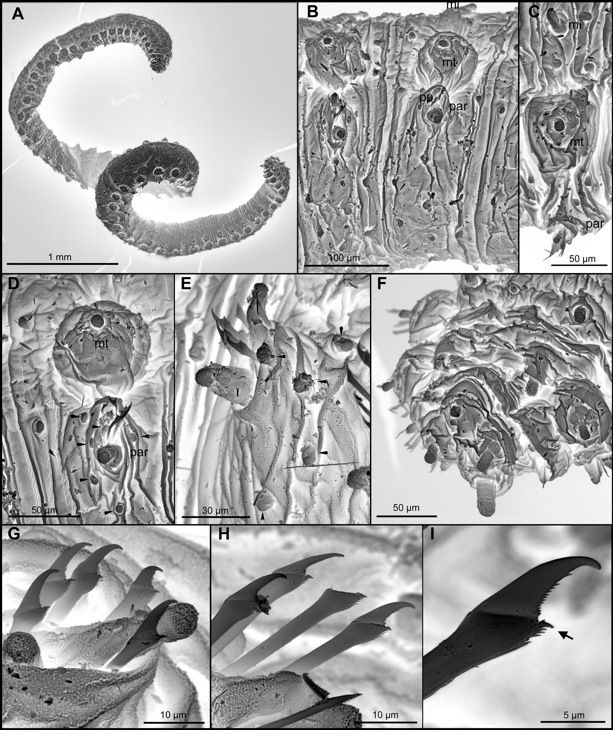
( Figs 2 View FIGURE 2 E, F, 4, 5, 6)
Type material: Holotype: AM W.47516, Australia, Queensland, Great Barrier Reef, beach near research station, 14°40'43"S, 145°26'50"E, intertidal, coarse sand, 5 Nov 2002. Paratypes: AM W.47517 (1 spec.), AM W.47518 (1 spec, on SEM stub), SIO-BIC A3645 (2 males and 2 females, in resin blocks), SIO-BIC A 3647 (2 specs), all same collection information as holotype.
Comparative material examined. Holotype of Sphaerodoropsis multipapillata heteropapillata Hartmann- Schröder, 1987, ZMH P.18875, Australia, Victoria, Geelong, Point Lonsdale, Abrasion terrace near lighthouse, on coralline algae, 24 Dec 1975. Holotype of Sphaerodoridium multipapillata Hartmann-Schröder, 1979 , ZMH P.14336, Tanzania, Mtwara, fine sand near coral reef south to the entrance of the harbour, 13 Nov 1967.
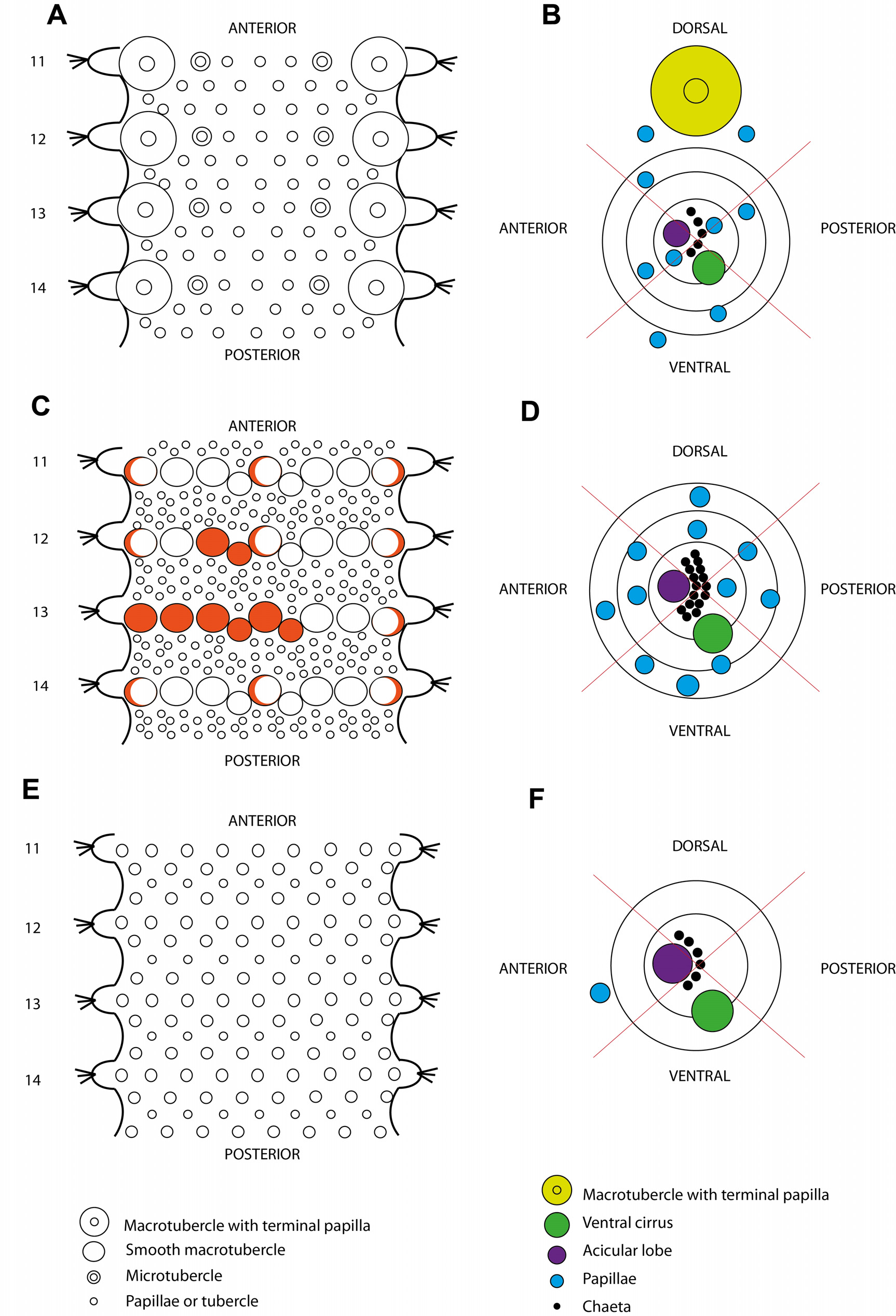
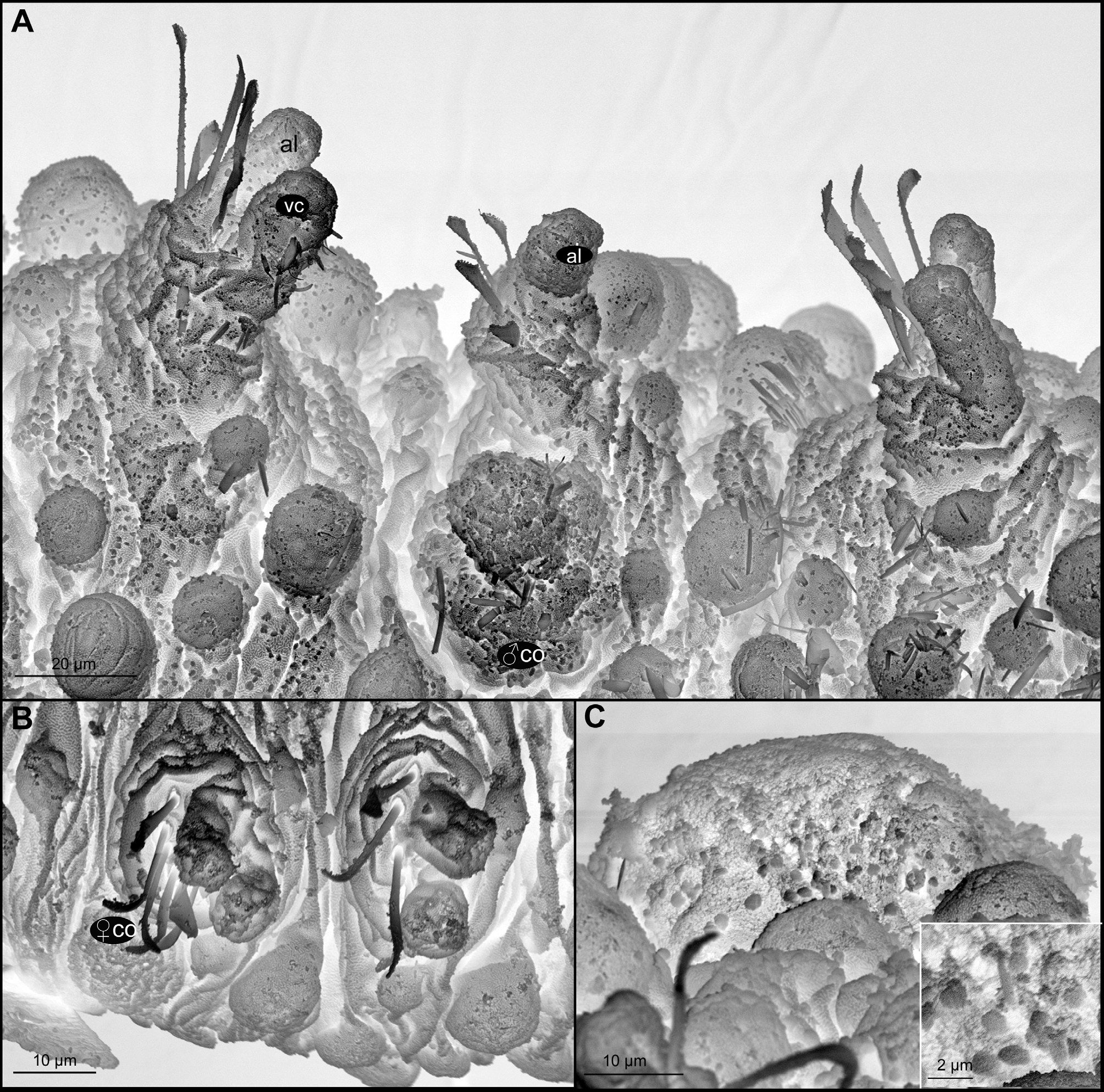
Diagnosis. Body ellipsoid, with strongly convex dorsum. Over 12 more or less clearly arranged longitudinal rows of spherical, variable in size, and sessile dorsal tubercles, in four transverse row per segment. Parapodia with digitiform acicular lobe similar in shape and size to ventral cirrus, lacking papillae. Around six semi-compound chaetae per parapodium with distally enlarged shaft and short blades (up to twice longer than wide), serrated, with distal long spines; shaft with conspicuous spinulation. Male with enlarged, bottle-shaped and porous copulatory organ present in chaetiger 6 ventral to each parapodia instead of the normal ventral cirri. Females with oval and flat tubercle with a porous surface also ventral to parapodia of chaetiger 6.
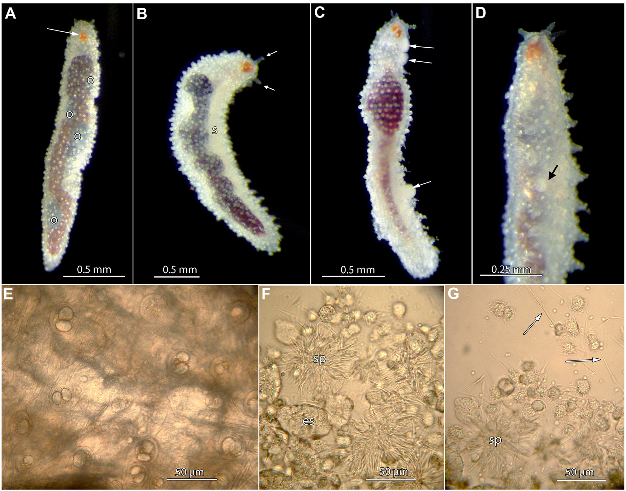
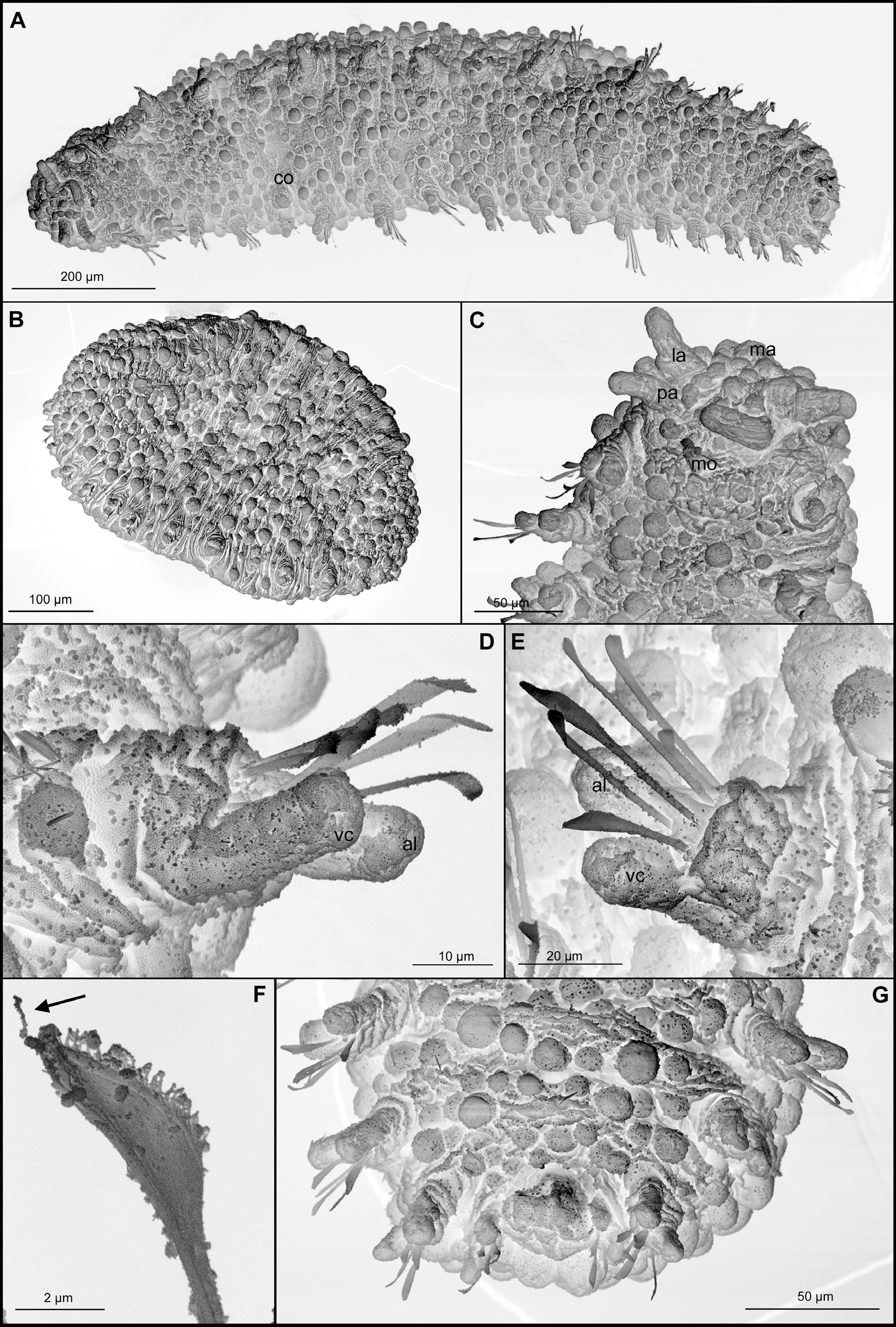
Description. Male holotype. Body more or less ellipsoid, anterior end bluntly rounded and posterior end tapering ( Figs 4 View FIGURE 4 A–D, 5A–B), measuring 1.5 mm long, 0.5 mm wide, with 18 chaetigers. Strongly convex dorsum and flat ventrum; segmentation inconspicuous ( Figs 4 View FIGURE 4 A–D, 5A–B). Live colour transparent with brown gut, milky white coelom; dark brown after preservation. Bright orange subdermal eyes ( Fig. 4 View FIGURE 4 A–D). Tegument with microscopic oblong granules. Head externally indistinct ( Fig. 5 View FIGURE 5 A–C). Prostomium with five appendages; a pair of ventral-most palps, digitiform and about three times as long as wide; a pair of lateral antennae similar in shape and size to palp; and a spherical median antenna ( Figs 4 View FIGURE 4 A–D, 5A). Around 10 small and spherical papillae confined by these appendages. A pair of tentacular cirri, shorter than palps and lateral antennae. Body surface covered in tubercles of two different sizes arranged in more or less 16–18 longitudinal rows in mid-body, four rows per segment, adding a total of around 30 in mid-chaetigers ( Figs 2 View FIGURE 2 E, 4A–D, 5B). Microtubercles absent. Ventrum with papillae similar in shape and size to dorsal, arranged in about 10 longitudinal rows, four transverse rows per segment and a total of about 20 papillae per chaetiger in mid-body ( Fig. 5 View FIGURE 5 A). Ventral papillae containing different type of granules ( Fig. 4 View FIGURE 4 E). Parapodial sub-conical, increasing in size towards chaetiger 3 and decreasing in last three chaetigers, as long as wide in mid-chaetigers ( Fig. 5 View FIGURE 5 A–E, G). Acicular lobe, digitiform, three times longer than wide, similar in shape as size as ventral cirrus ( Fig. 5 View FIGURE 5 D–E). Parapodia lacking parapodial papillae; but one spherical papilla located close to anterior base of each parapodia ( Figs 2 View FIGURE 2 F, 5D). Bottle-shaped copulatory organ present in chaetiger 6 ventral to each parapodia, which lack ventral cirri ( Figs 4 View FIGURE 4 D, 5A, 6A), suggesting these structures are transformed ventral cirri. Usually six chaetae arranged in a more or less curved transverse line behind the acicular lobe ( Fig. 5 View FIGURE 5 D–E). Hooks in anterior segments not observed. All chaetae simple ( Fig. 5 View FIGURE 5 F), appearing semi-compound in some cases, denoting that shaft and blade have probably fused. Distal end enlarged, three times longer than wide, with spinulation in cutting edge and a conspicuous distal projection ( Fig. 5 View FIGURE 5 F), similar along body and within fascicles. Chaetae showing a thin groove running parallel to edge, in distal end. Pygidium terminal, with inconspicuous spherical single anal cirrus ( Fig. 5 View FIGURE 5 G). Brown gut seen by transparency, coiled in some parts ( Fig. 4 View FIGURE 4 A–D) without a distinct muscular structure.
Variation. Paratypes range from 0.6 to 1.5 mm long, and 12–20 segments, showing two different body shapes, more or less elongated, probably due to contraction. The length of the anterior appendages also varies, being contracted in one paratype and resembling prostomial papillae ( Fig. 5 View FIGURE 5 B), while in rest of paratypes they are digitiform and conspicuous. Number and size of epithelial tubercles vary slightly along chaetigers but they are constantly arranged in about four dorsal and ventral more or less evident transverse rows per segment. Longitudinal rows range between 13–18, however numbers are often difficult to count due to the different size of tubercles and their zigzag arrangement. Papillae seem absent of parapodia in all specimens, however and as mentioned in the holotype, an anterior ventral papillae can be considered part of the parapodia when these are relaxed and enlarged. Chaetae often broken but those that are intact are as those described in holotype, simple with spinulation along cutting edge and a distal spine. All specimens (live and preserved) lack external pigmentation.
Male paratypes share the presence of copulatory organs as described in holotype, and ventral to parapodia of chaetiger 6 ( Fig. 6 View FIGURE 6 A). Females bear an oval and flat tubercle with a porous surface also ventral to parapodia of chaetiger 6 ( Fig. 6 View FIGURE 6 B). Females, instead, show ventral cirri on the parapodia of this chaetiger ( Fig. 6 View FIGURE 6 B).
Remarks. The present species agrees with the diagnostic features attributed to those Sphaerodoropsis species with macrotubercles in more than two transverse rows per chaetigers (Group 3, according to Borowski 1994), but also with other genera considered to lack macrotubercles and with large papillae instead, such as Amacrodoum Kudenov 1987, Commensodoum Lützen, 1961 and Euritmia Sardá-Borroy, 1987 . Distinguishing papillae and macrotubercle is imprecise since they are currently defined according to their size ( Fauchald 1974) and this is continuous character. Consequently, the species sharing the presence of more than 10 longitudinal rows of tubercles (more or less similar in size) in around four transverse rows per chaetiger need revision to enlighten their relationships and address their correct classification (Capa & Bakken in prep.).
There are two other species of Sphaerodoropsis in Borowski’s Group 3, S. multipapillata (Hartmann-Schröder, 1974) , from Tanzania, and S. mutipapillata heteropapillata Hartmann-Schröder, 1987 , from Victoria, Australia, recently given a species rank (Capa & Bakken 2015). Sphaerodoropsis plurituberculata n. sp. is most similar to S. heteropapillata because both species are covered with dissimilar dorsal tubercles, while in S. multipapillata these are of similar size ( Hartmann-Schröder 1974b; Hartmann-Schröder 1987; Capa & Bakken, 2015). Differences between S. heteropapillata and the new species include the length of the prostomial appendages (inconspicuous in S. heteropapillata and digitiform in S. plurituberculata n. sp., at least when not contracted); the number and of ventral papillae (around 50 per segments in S. heteropapillata and 30 in S. plurituberculata n. sp.); and the number of parapodial papillae ( S. heteropapillata bears one at anterior surface while S. plurituberculata n. sp. lacks parapodial papillae).
Amacrodorum bipapillatum Kudenov, 1987 , from Alaska, is distinguished from the new species by the morphology of the antennae, with spurs in A. bipapillatum and apparently smooth in the new species; the presence of one pair of black eyes in A. bipapillatum , absent in the new species; and having chaetae with strongly recurved tips ( Kudenov 1987b: Fig. 1 View FIGURE 1 J) instead of straight but provided with a long and thin distal spine. Moreover, A.
bipapillatum was described with the apparently atypical attribute of bearing two different types of tubercles, elliptical and hemispherical while in S. plurituberculata n. sp. all papillae seem spherical.
Euritmia hamulisetosa Sardá-Borroy, 1987 View in CoL described from the south of Spain, differs from the new species in the chaetal morphology. Euritmia hamulisetosa View in CoL bears simple chaetae, of different widths in all chaetigers all provided with a distal spine bent rearward to the cutting edge ( Sardá-Borroy 1987) while S. plurituberculata n. sp. bears semi compound chaetae, all similar in width and with an erected thin spine. The other congener, Euritmia capense ( Day, 1963) View in CoL is provided with two defined rows of papillae of different size each, but they are not distributed in an apparently random pattern as in S. plurituberculata n. sp.
Commensodorum commensalis Lützen, 1961 View in CoL is distinguished from the new species in the presence of four longitudinal rows of papillae along the dorsum ( Lützen 1961).
Reproductive notes. Live specimens of Sphaerodorpsis plurituberculata n. sp. maintained in aquaria were studied during several days. White masses were observed attached to the body surface of both sexes during this time. These were found to be spermatophores when examined with a compound microscope (image not shown).
On several occasions males were seen to be pseudo-copulating with females, or other males, for less than a minute and on detaching were seen to have left one or two spermatophores on the body surface ( Fig. 4 View FIGURE 4 C). Masses that corresponded to the size and colour of these spermatophores could be seen ventrally in chaetiger 6 ( Fig. 4 View FIGURE 4 D), suggesting this segment is indeed the location of copulatory papillae. Structures attributed to be copulatory organs have been described for several other sphaerodorids in Sphaerodoropsis View in CoL and Sphaerodoridium View in CoL (Moreira et al. 2004; Böggemann 2009; Reuscher & Fiege 2011; Moreira & Parapar 2012, 2015; Capa & Bakken 2015). Males of Sphaerodoropsis plurituberculata n. sp. were observed to have various stages of spermiogenesis in their coelom. Sperm developed in cluster attached to a large central cytophore ( Fig. 4 View FIGURE 4 F–G). The sperm heads were elongate at ~35 µm long ( Fig. 4 View FIGURE 4 G) with what appeared to be a free flagellum of similar length. Free sperm were visible in the coelom ( Fig. 4 View FIGURE 4 B) and it is not known how the sperm were packaged in the spermatophores. Males could be easily distinguished from females by having milky white coelomic contents surrounding the dark gut. Females had bluish/ gray oocytes at various stages of development, with up to 10 oocytes at what appeared to be a late stage of development ( Fig. 4 View FIGURE 4 A). Some females were observed to have free sperm in the coelom that had presumably come from the spermatophores (not shown). The largest eggs observed inside a female were about 150 µm in diameter suggesting development is lecithotrophic, similarly to conclusions reached by Christie (1984).
| ZMH |
Zoologisches Museum Hamburg |
No known copyright restrictions apply. See Agosti, D., Egloff, W., 2009. Taxonomic information exchange and copyright: the Plazi approach. BMC Research Notes 2009, 2:53 for further explanation.
|
Kingdom |
|
|
Phylum |
|
|
Class |
|
|
Order |
|
|
Family |
|
|
Genus |
Sphaerodoropsis plurituberculata
| Capa, María & Rouse, Greg W. 2015 |
Euritmia hamulisetosa Sardá-Borroy, 1987
| Sarda-Borroy 1987 |
Euritmia capense (
| Day 1963 |
Commensodorum commensalis Lützen, 1961
| Lutzen 1961 |