
Echinohelea Macfie
|
publication ID |
https://doi.org/10.11646/zootaxa.3879.1.1 |
|
publication LSID |
lsid:zoobank.org:pub:6423894B-97D9-4286-ABB9-D4AF072B57FD |
|
DOI |
https://doi.org/10.5281/zenodo.5593009 |
|
persistent identifier |
https://treatment.plazi.org/id/027587C9-BD67-303C-FD66-18314C72E0AC |
|
treatment provided by |
Felipe |
|
scientific name |
Echinohelea Macfie |
| status |
|
Echinohelea Macfie View in CoL View at ENA
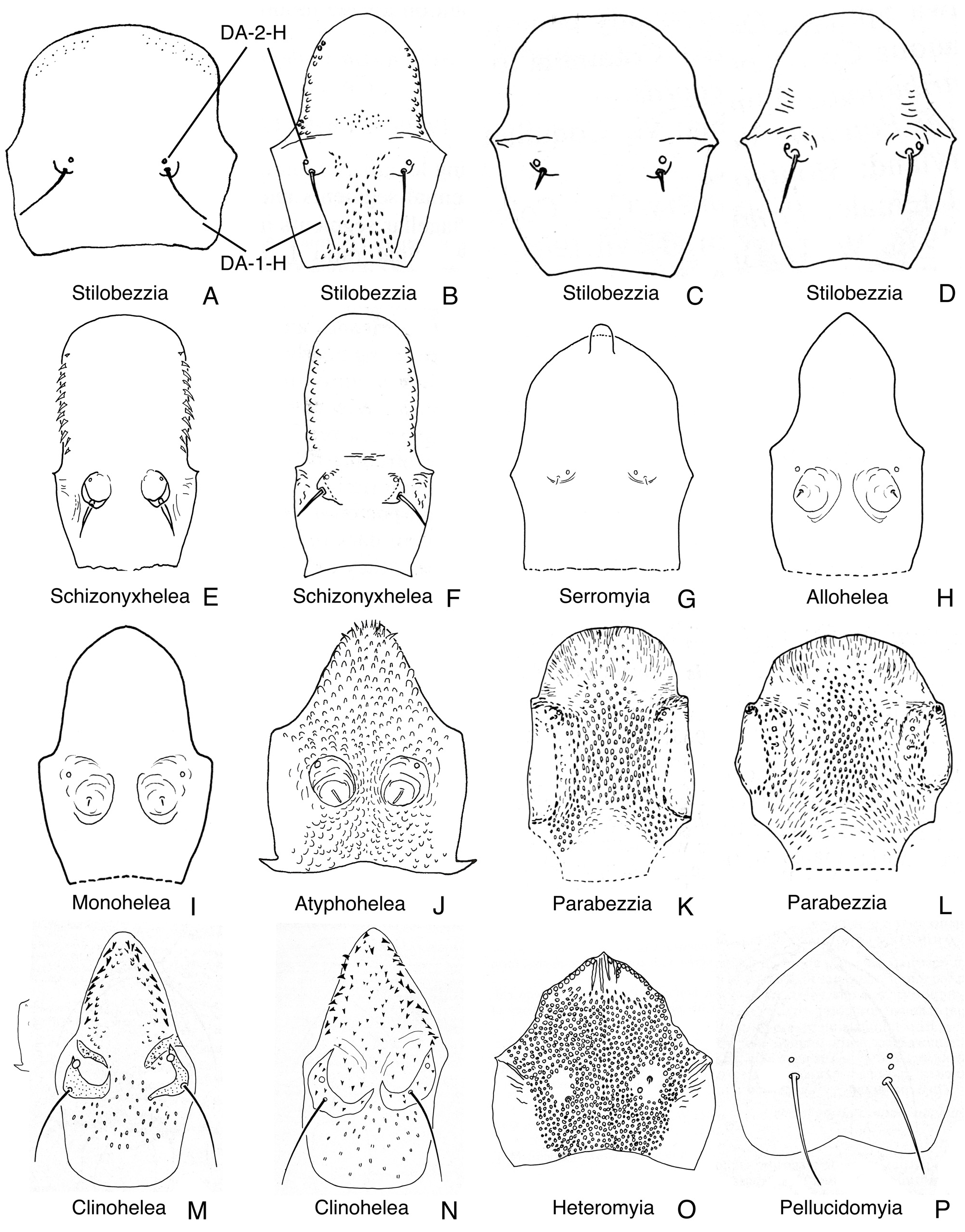
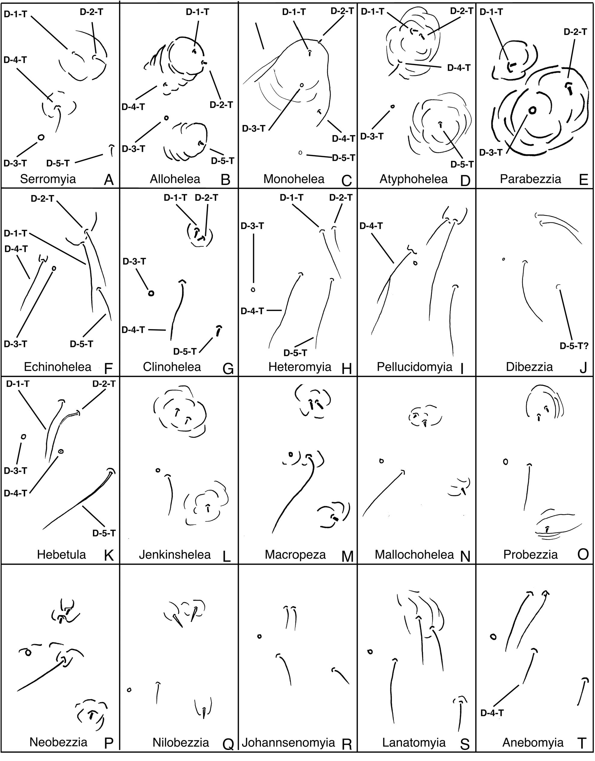
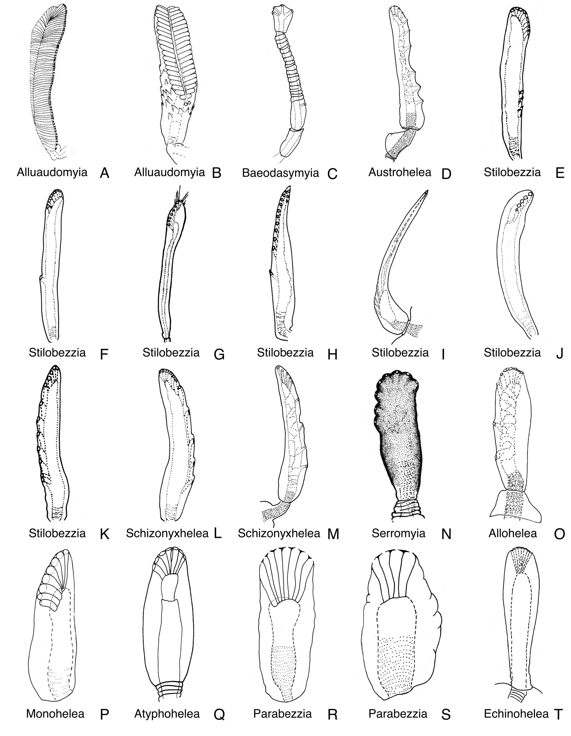
( Figs. 20 View FIGURE 20 , 26A View FIGURE 26 , 30F View FIGURE 30 , 31K View FIGURE 31 , 37C View FIGURE 37 , 44T View FIGURE 44 , 50C View FIGURE 50 , 63A View FIGURE 63 , 75B View FIGURE 75 )
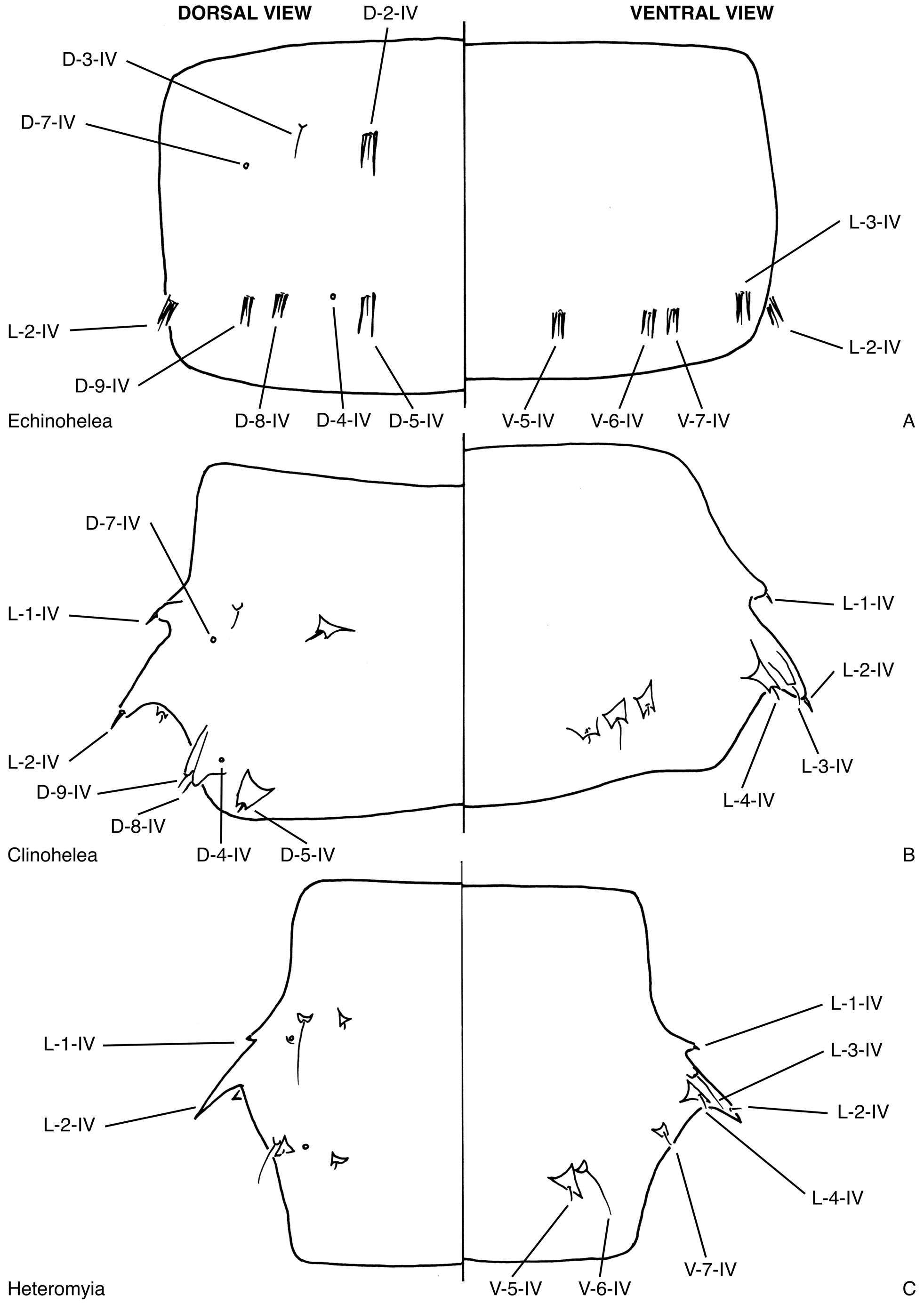
DIAGNOSIS: Only pupa of Ceratopogonidae with abdominal segment 4 with only two lateral sensilla (L-2-IV, L- 3-IV) and with most sensilla at the bases of bifid tubercles ( Fig. 63A View FIGURE 63 ).
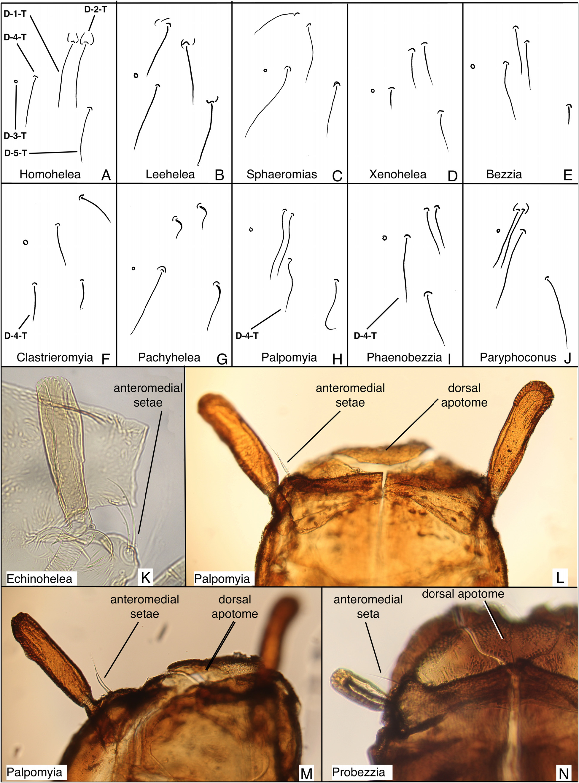
DESCRIPTION: Total length = 2.91 mm. Without larval exuviae retained on abdomen. Exuviae with flagellum appressed against lateral margin of face, midleg, wing (as in Figs. 16B View FIGURE 16 , 33B View FIGURE 33 ). Ecdysial tear uncertain but not around base of antenna; at least medial to antennal base (as in Figs. 15B View FIGURE 15 , 79D View FIGURE 79 ). Head: Dorsal apotome missing; dorsolateral cephalic sclerite (as in Fig. 13H View FIGURE 13 ) fused to scutum, each side uncertain in whole pupa; mouthparts ( Fig. 26A View FIGURE 26 ) with mandible well-developed, lacinia absent; palpus extending posterior to posterolateral margin of labium; labium separated medially by labrum, hypopharynx; apex of antenna ( Fig. 37C View FIGURE 37 ) anterior to posterior extent of midlength portion of midleg (portion lateral to mesosternum), narrowed posteriorly; sensilla: dorsal apotomals ( Fig. 20 View FIGURE 20 )—uncertain; dorsolateral cephalic sclerite sensilla—1 elongate seta, 1 campaniform sensillum; clypeallabrals ( Fig. 26A View FIGURE 26 )—1 small seta; oculars ( Fig. 26A View FIGURE 26 )—1 campaniform sensillum. Thorax: Prothoracic extension ( Fig. 26A View FIGURE 26 ) wide, well-developed, extending from palpus to antenna; mesonotum with short tubercles, extending posteriorly but medial division of metathorax uncertain ( Fig. 50C View FIGURE 50 ); respiratory organ ( Fig. 44T View FIGURE 44 ) length/width = 4.71, elongate, slender, somewhat flattened, at least apically (perhaps more), with pores closely abutting at apex of respiratory organ, arranged in single row, outer surface smooth, with short, wide pedicel, base with elongate posteromedial apodeme, membranous base of respiratory organ moderately elongate, tracheal tube straight to slightly curved along length, surface smooth; wing ( Fig. 37C View FIGURE 37 ) with apical tubercle lateral to apex of hind leg, separated medially by fore-, midlegs; halter apex and hind leg (as in Fig. 33A View FIGURE 33 ) narrowly abutting; halter apex abutting anterolateral knob-like extension of tergite 2; legs ( Fig. 37C View FIGURE 37 ) with lateral margin of foreleg near midlength of wing evenly curved; hind leg visible at lateral margin of wing (as in Fig. 32L View FIGURE 32 ); with apex of foreleg slightly anterior to apex of midleg; apex of hind leg abutting apex of midleg laterally; sensilla: anteromedials—2 elongate setae, 1 campaniform sensillum ( Fig. 31K View FIGURE 31 ); anterolaterals—1 moderately long seta; dorsal setae ( Fig. 30F View FIGURE 30 )—D-1- T, D-2-T, D-4-T, D-5-T setae, D-3-T campaniform sensillum; D-1-T, D-2-T on single tubercle, D-3-T posteromedial to D-4-T; supraalar 2—campaniform sensillum; metathoracics ( Fig. 50C View FIGURE 50 )—1 seta on one side only, 2 campaniform sensilla on both sides; M-3-T near anterior margin of metathorax. Abdomen: without pigmentation pattern, segment 2 as wide or slightly wider than segment 3, segments with undivided, thin to thick setae, with rounded to bilobed, short to somewhat elongate tubercles, tergites or sternites entire, each without membranous disc; segment 9 ( Fig. 75B View FIGURE 75 ) not strongly modified, terminal processes closely approximated basally, each projecting posterodorsolaterally, tapering to pointed apex; sensilla: tergite 1 ( Fig. 50C View FIGURE 50 ) with 7 setae, 2 campaniform sensilla, including 3 lateral sensilla, D-2-I, D-3-I closely approximated, D-7-I situated anteriorly near D-3-I; segment 4 ( Fig. 63A View FIGURE 63 )—D-2-IV short seta on bifid tubercle; D-3-IV moderately elongate seta without tubercle; D-5-IV, D-8-IV, D- 9-IV short setae; at base on bifid, separate tubercles, posterior dorsal sensilla in transverse row, arranged medially to laterally: D-5-IV, D-4-IV, D-8-IV, D-9-IV; D-7-IV situated anterorlaterally near D-3-IV; L-1-IV absent, L-2-IV, L-3-IV short setae each at base of bifid tubercle, L-4-IV absent, V-5-IV, V-6-IV, V-7-IV moderately elongate setae, at base of bifid, slender tubercles, V-6-IV, V-7-IV closely approximated; segment 8 without D-3-VIII, without L-1- VIII; segment 9 ( Fig. 75B View FIGURE 75 )—with D-5-IX, D-6-IX campaniform sensilla.
DISTRIBUTION AND HABITAT: The genus Echinohelea is known from 27 species from nearly every Region worldwide but is absent from the Palaearctic Region ( Borkent 2014). An unpublished record from Borneo is known (pers. obs.). The only known immature, the pupa of E. lanei described here, was collected from Flanders, New York, USA. The Flanders Pond site was scrubby red maple growth flooded with a few inches of clear water (Hugo Jamnback, pers. comm.). Grogan (1975) reported a reared specimen of E. lanei collected from under bark of a rotting log in Maryland, USA and Knausenberger (1987) found this species in damp dark brown mud (his collection No. 29) in a roadside ditch in Virginia, USA. De Meillon & Wirth (1991) noted two African species reared from wet soil and from mud at a pond margin.
TAXONOMIC DISCUSSION: Only one moderately well preserved pupa of Echinohelea is known, that of E. lanei ( Tables 2–3 View TABLE 2 View TABLE 3 ). Wirth (1994b) provided an earlier description. Details of the sensilla distribution of abdominal segment 4 differs here because the specimen was distorted by compression and is reconstructed here. The dorsal apotome of the specimen was not present and cannot, therefore, be characterized.
Wirth (1994b) noted that the pupa of Echinohelea was similar to some Heteromyiini (as described by Elson-Harris & Kettle 1986) and this is reflected in the phylogenetic relationship supported here (and Borkent 1995) between Echinohelea and Heteromyiini + Sphaeromiini s. lat. + Palpomyiini + Stenoxenini .
MATERIAL EXAMINED: E. lanei : 1 pupal exuviae, Flanders Pond, Flanders, New York, USA, 4-VI-1963 (NYSM).


No known copyright restrictions apply. See Agosti, D., Egloff, W., 2009. Taxonomic information exchange and copyright: the Plazi approach. BMC Research Notes 2009, 2:53 for further explanation.
|
Kingdom |
|
|
Phylum |
|
|
Class |
|
|
Order |
|
|
Family |
|
|
SubFamily |
Ceratopogoninae |
|
Tribe |
Ceratopogonini |