
Nanophareus bipartitus, Hara, Marcos Ryotaro, Pinto-Da-Rocha, Ricardo & Kury, Adriano Brilhante, 2012
|
publication ID |
https://doi.org/ 10.5281/zenodo.212388 |
|
DOI |
https://doi.org/10.5281/zenodo.6175470 |
|
persistent identifier |
https://treatment.plazi.org/id/3C3487A8-426E-9D32-DDEB-B08FFED531AC |
|
treatment provided by |
Plazi |
|
scientific name |
Nanophareus bipartitus |
| status |
sp. nov. |
Nanophareus bipartitus View in CoL sp. nov.
( Figs. 6 View FIGURE 6 , 7 View FIGURE 7 , 11 View FIGURE 11 A–B, 12C–D, 13)
Type material. CHILE. V Región de Valparaíso: Parque Nacional La Campana (32°58´48”S, 71°07´03”W), 16.I.2010, R. Pinto-da-Rocha, F. Cádiz L. & D. Cádiz L. leg., ma holotype (MNHNCL); idem, 1 ma & 1 fe paratypes ( MZSP 43034); idem, (Sector Granizo, 32°58´30”S, 71°07´36”W), 20.XII.2009, L. Almeida et al. leg., 1 ma paratype ( IBSP 10543). Additional material: CHILE. V Región de Valparaíso: Parque Nacional Los Cipreses (34°17´40”S, 70°26´50”W), 17.I.2010, R. Pinto-da-Rocha, F. Cádiz L. & D. Cádiz L. leg., 1 fe exoskeleton without appendages ( MZSP 43035).
Diagnosis for males. Nanophareus bipartitus sp. nov. resembles N. palpalis because of the unarmed frontal hump on dorsal scutum, widened ocularium, unarmed scutal area III, prolateral apical apophysis of coxa IV barely reaching the posterior margin of dorsal scutum, trochanter IV unarmed prolateral apically and femur IV without retromedian apophysis. Nanophareus bipartitus sp. nov. can be distinguished from N. palpalis by: Scutal area IV divided by a longitudinal median groove and tibia IV unarmed retrolaterally, with two ventral rows of tubercles increasing in size apicad, 2 ventral apical blunt spines. Nanophareus bipartitus sp. nov. can be distinguished from the other species of the genus by the: Coxa IV with single-branched prolateral apical apophysis; and scutal area IV divided by a longitudinal median groove.
Diagnosis for females. Nanophareus bipartitus sp. nov. can be distinguished from the other species of the genus by the combination of the following characters: Ocularium widened; ocularium and frontal hump unarmed; and scutal area IV divided by a longitudinal median groove.
Etymology. In reference to scutal area IV divided by a longitudinal median groove, an unusual feature among Pachylinae .
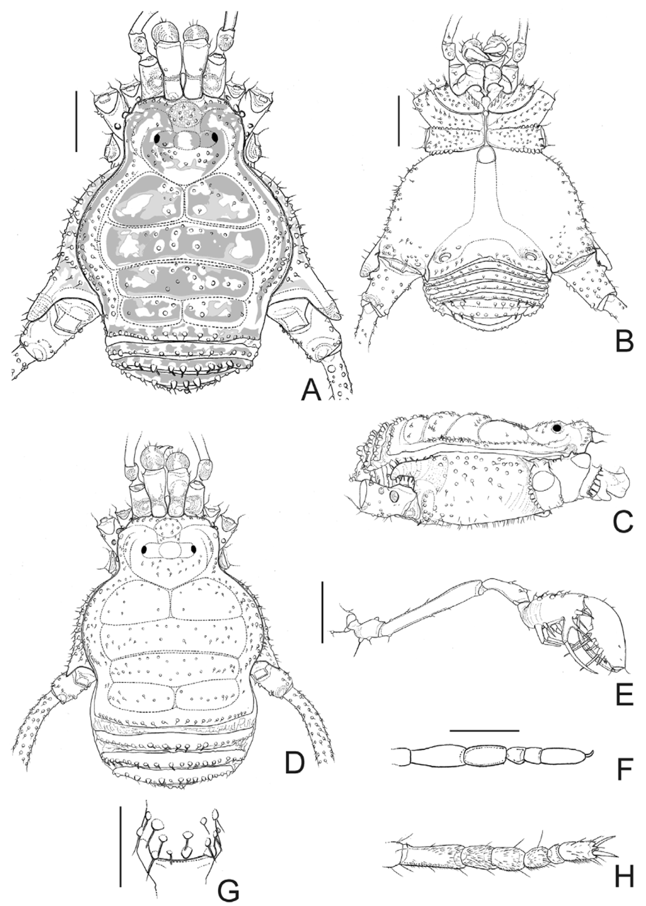
Description. Male (holotype MNHNCL): Dorsum ( Fig. 6 View FIGURE 6 A, C): Measurements: DSL 3.65; DSW 3.35; LI 8.10; LII 18.00; LIII 10.35; LIV 17.75. Median tuberculate frontal hump, with 4–5 tubercles on each side of anterior margin of carapace. Ocularium widened, low, with median eminence, 1 tubercle near each eye. Carapace with sparse tubercles. Scutal area I with most of 15 tubercles near median groove and groove I; II with 12 tubercles concentrated the middle; III with 13 scattered tubercles; IV divided by a longitudinal median groove, with 4–5 tubercles in each half. Lateral margin of dorsal scutum with a row of enlarged tubercles inserted among small ones, more densely distributed between grooves II and IV. Posterior margin of dorsal scutum and free tergites I–III each one with a row of 18, 15, 13, 10 tubercles, respectively, those median ones in free tergite III largest. Anal operculum with 12 tubercles on median region and small tuberculate on its posterior margin.
Venter ( Fig. 6 View FIGURE 6 B): Coxa I–III densely tuberculate; IV with tubercles scattered on the sides, stigmatic area and surrounding region almost smooth. Posterior margin of stigmatic sternite and free sternites each one with a row of tubercles. Anal operculum tuberculate.
Chelicera: Segment I with 1 tubercle, bulla weakly marked; movable finger with 4 teeth; fixed finger with 4 teeth.
Pedipalpus ( Fig. 6 View FIGURE 6 E): Coxa with 1 ventral tubercle, dorsally smooth. Trochanter with 1–2 dorsal tubercles, 2 ventral tubercles. Femur with few small tubercles. Patella and tibia dorsally tuberculate. Tibial setation: Prolateral IIiII, retrolateral Ii[Ii]i (subapical bifid and longest, 1 short and 1 long setae). Tarsal setation: Prolateral IiIii, retrolateral iIiIii, iIiIIii.
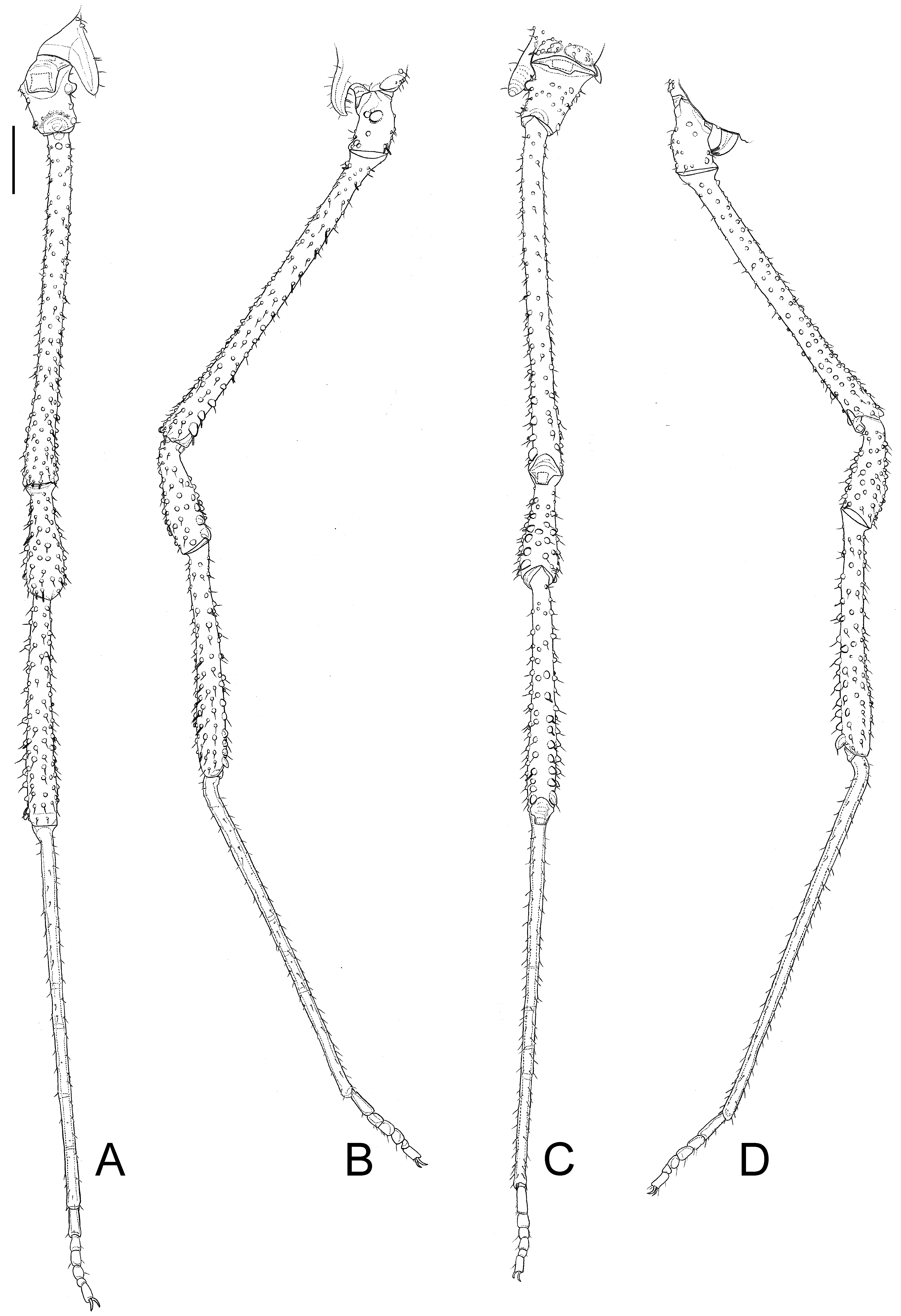
Legs ( Figs. 6 View FIGURE 6 F–H, 7): Coxa IV densely tuberculate, with prolateral apical apophysis single-branched, directed ventrally, a short, conical retrolateral apical apophysis. Trochanters I–IV tuberculate; IV 1.5 times longer than wide, prolaterally with a short, conical, blunt median apophysis, swollen in the middle; retrolaterally with 2 basal, 1 apical enlarged tubercles. Femur IV straight, long, dorsally unarmed; ventrally with the prolateral row of tubercles increasing in size subapically, 1 prolateral apical blunt spine, 1 retrolateral apical slightly enlarged tubercle. Patella IV tuberculate, proventral ones enlarged, 1 proventral apical blunt spine. Tibia IV ventrally with two rows of tubercles increasing in size distad, retrolateral ones enlarged, 2 apical blunt spines. Basitarsus I slightly swollen. Tarsal process reduced to a seta. Tarsal segmentation: 5(3); 9(3); 6; 6.
Penis ( Fig. 11 View FIGURE 11 A–B): Glans with wide sac and a small dorsal projection; stylus slender, cylindrical, curved with scattered ventral median trichomes; ventral process with nail head-like apex (apex with lateral projections) directed to stylus. Ventral plate distal setae slightly curved on apex, placed almost on ventral plate corner; ventral plate basal setae slightly curved on apex (larger than distal group).
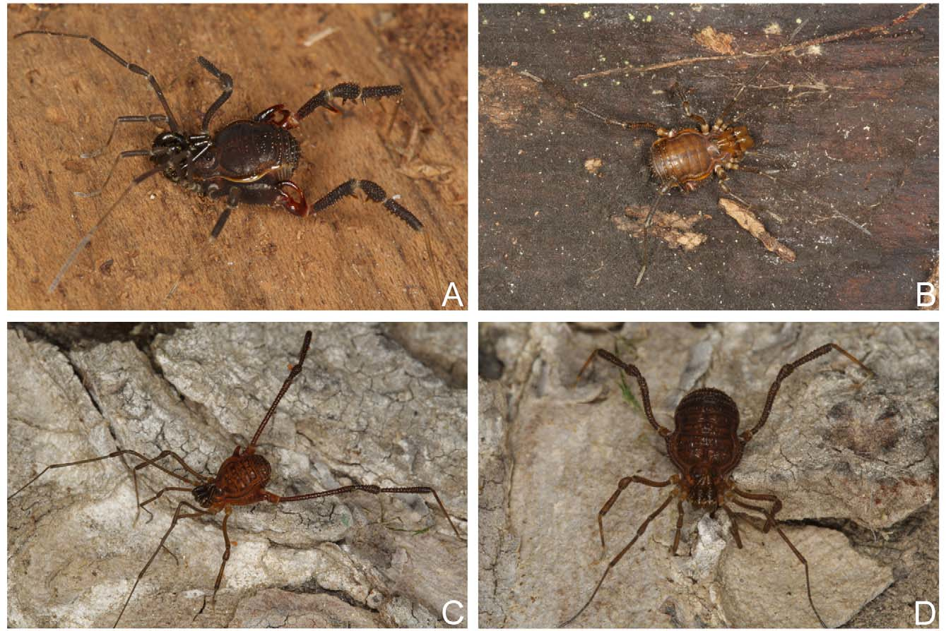
Coloration in live specimen ( Fig. 12 View FIGURE 12 C): Scutal areas, posterior margin of dorsal scutum and free tergites light brown with patches of black pigment; lateral margin of dorsal scutum with one narrow, almost black stripe. Chelicera, pedipalp and legs greenish background with dark brown reticulated pattern. Apex of apophyses of coxa IV and basal half of trochanter IV orange.
Female (paratype; MZSP 43034): Dorsum ( Figs 6 View FIGURE 6 D, 12D): Measurements: DSL 3.35; DSW 2.90; LI 5.65; LII 9.20; LIII 6.85; LIV 9.25. Scutal area III with one irregular row of 11 tubercles, one posterior row with 15 tubercles. Pedipalpus: Tibial setation: Prolateral IiIi, retrolateral i[Ii]i, Ii[Ii]i (subdistal setae bifid). Tarsal setation: Prolateral IiIi, retrolateral iIiIi, iIiIii. Femur IV slightly curved inwards, with proventral row of enlarged tubercles on distal ¼, ventroapical as in male. Patella–tibia IV with tubercles slightly increasing in size apically, patella IV with a proventral apical blunt spine and tibia IV with two apical enlarged tubercles, retroventral one largest. Tarsal segmentation: 5(3); 6–7(3); 6; 6.
Variation in males (n=3): Measurements: DSL 3.60–3.70; DSW 3.10–3.35; LI 7.95–8.50; LII 17.25–18.80; LIII 10.35–10.50; LIV 16.65–17.75. Pedipalpus: Femur with 1–2 prolateral subapical seta(e); tibial setation: Prolateral IIii, IIiI, IIiIi, IIiII, retrolateral Ii[Ii]i; tarsal setation: Prolateral IiIi, IiIii, retrolateral IiIii, iIiIii, iIiIiii, iIiIIii. Tarsal segmentation: 5–6(3); 8–9(3); 6; 6.
Geographical distribution ( Fig. 13 View FIGURE 13 ): Central Chile. Valparaíso.
Biotope note: The type locality of Nanophareus bipartitus and N. araucanus , the Sector Granizo of Parque Nacional La Campana, is in west slope of Andean Cordillera, in low altitude (about 400–500 m), in sclerophyllous forest in Mediterranean climate. It receives about 480 mm during winter (May to August) and 120 mm from September to April ( Mooney 1977; CONAF 1982).


No known copyright restrictions apply. See Agosti, D., Egloff, W., 2009. Taxonomic information exchange and copyright: the Plazi approach. BMC Research Notes 2009, 2:53 for further explanation.
|
Kingdom |
|
|
Phylum |
|
|
Class |
|
|
Order |
|
|
Family |
|
|
Genus |