
Terebellides longiseta, Parapar & Martin & Moreira, 2020
|
publication ID |
https://doi.org/ 10.11646/zootaxa.4771.1.1 |
|
publication LSID |
lsid:zoobank.org:pub:A46FAF72-6F95-4DA3-A41D-FE770D6EDF1F |
|
DOI |
https://doi.org/10.5281/zenodo.3816159 |
|
persistent identifier |
https://treatment.plazi.org/id/33AEE210-CA77-488F-A43B-2FDC23375CBE |
|
taxon LSID |
lsid:zoobank.org:act:33AEE210-CA77-488F-A43B-2FDC23375CBE |
|
treatment provided by |
Plazi |
|
scientific name |
Terebellides longiseta |
| status |
sp. nov. |
Terebellides longiseta sp. nov.
Figures 11C View FIGURE 11 , 21B View FIGURE 21 , 25−27 View FIGURE 25 View FIGURE 26 View FIGURE 27 , 37 View FIGURE 37 ; Tables 1, 2 urn:lsid:zoobank.org:act:33AEE210-CA77-488F-A43B-2FDC23375CBE
Material examined. Type material. Holotype (NHMD-231459); paratypes: five specimens (NHMD-231437, NHMD-231448 and NHMD-636926). Non-type material. One specimen ( MNCN 16.01 View Materials /18591, Table 1) .
Diagnosis. Body medium/big sized (up to 21 mm in length). Anterior branchial lobe large; posterior ventral lobes thinner than dorsal ones, with long terminal filament, directly emerging from branchial stem. TC1 about as long as following ones; all thoracic notochaetae very long and numerous, with rostrum vs capitium length of about 1/1, and capitium with a first row of 3 big teeth followed by much smaller ones.
Description based on holotype
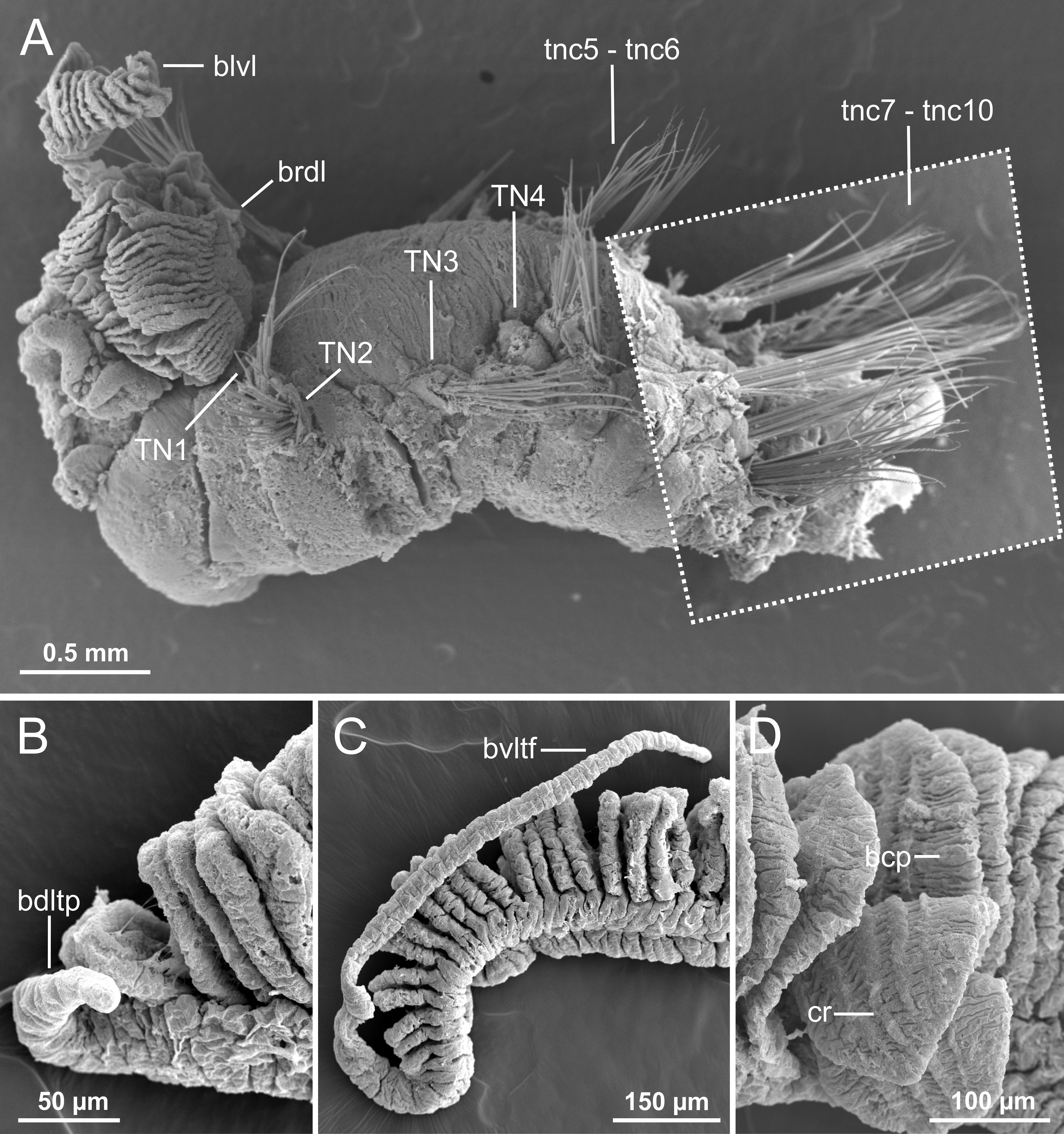
Measurements and general body features. Incomplete specimen, broken at TC10; 8.0 mm long and 3.5 mm wide ( Fig. 21B View FIGURE 21 , 25A View FIGURE 25 ). Prostomium compact; large tentacular membrane surrounding mouth; buccal tentacles lost. SGI as an expanded structure below tentacular membrane. Lateral lappets on SG3–6 (TC1–4), especially large in SG3 (TC1) forming a fan-shaped antero-dorsal expansion ( Fig. 25 View FIGURE 25 B–C, arrow).
Branchiae. Branchiae arising as single structure from SG3, with a single stalked mid-dorsal branch having one pair of dorsal (upper) lobes (1+2), not fused along their length, and one pair of ventral (lower) lobes (3+4) of similar length but much thinner, neither fused together nor to dorsal ones ( Fig. 21B View FIGURE 21 , 25B, D View FIGURE 25 ). Anterior lobe (lobe 5) well developed, triangular, about half to 1/3 length of posterior dorsal lobes ( Fig. 25C View FIGURE 25 ). Pointed projection of both upper and lower posterior lobes much longer in ventral ones, becoming a long terminal filament ( Fig. 25B, D View FIGURE 25 , 26 View FIGURE 26 B−C). Both sides of branchial lamellae with several parallel rows of cilia and low ciliated tufts, giving a zig-zag appearance to outer edge of dorsal posterior lobe branchial lamellae under stereomicroscope ( Fig. 25B, D View FIGURE 25 ).
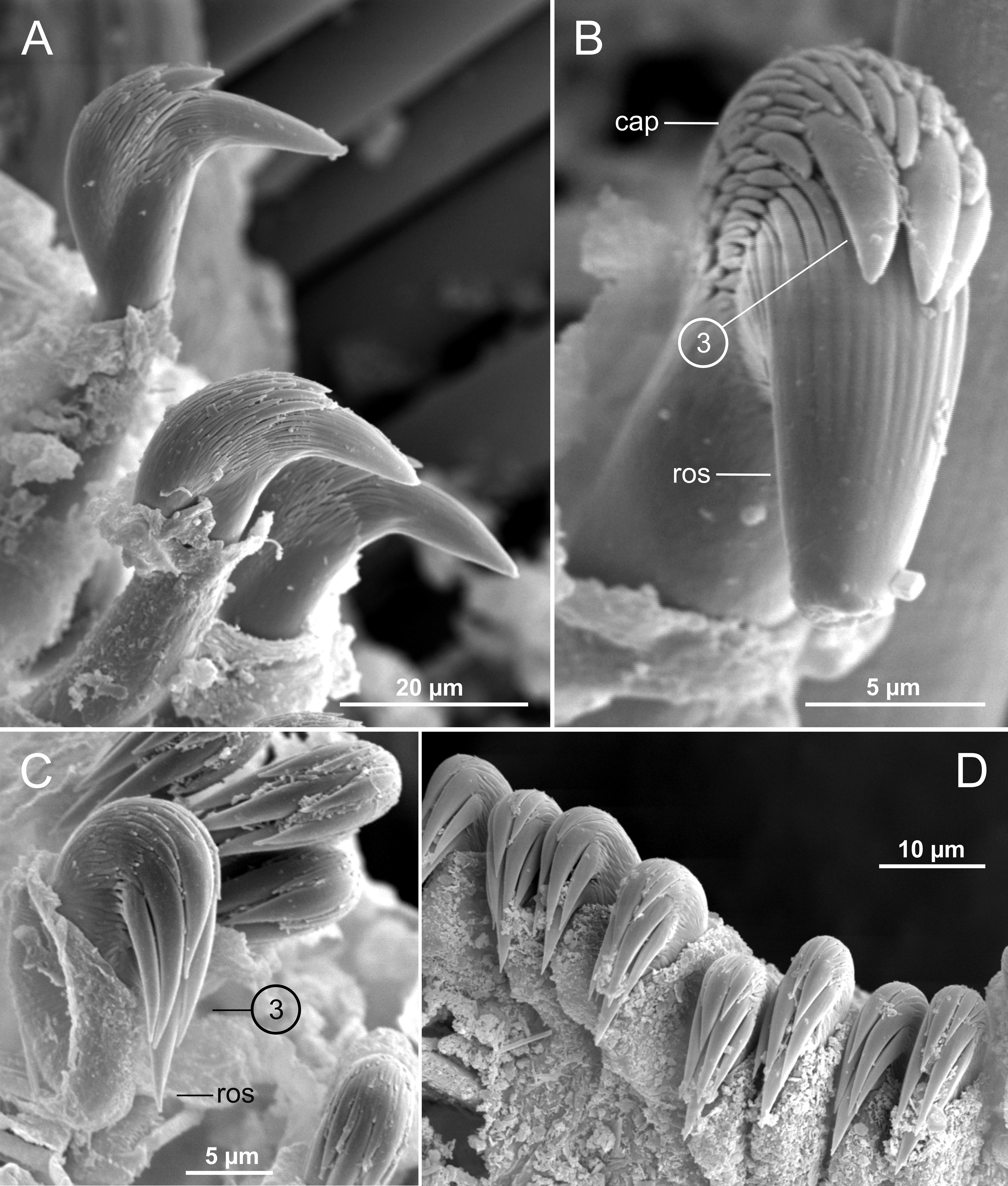
Thorax. Incomplete, with ten pairs of notopodia from SG3 to SG12 (TC1 to TC10). TC1 notopodia well developed, as long as subsequent ones ( Fig. 25 View FIGURE 25 B−C, 26A). All notochaetae simple capillaries with textured surface, very long (near body width at same level) and numerous (12–15 per notopodium). Neuropodia as sessile pinnules from TC6 (SG8), with uncini in single rows starting from TC7 (SG9) throughout. First neuropodia (TC6/TU1) with 8–9 sharply bent, acute-tipped, geniculate chaetae, having minute teeth forming a capitium visible under SEM. From TC7, neuropodia with about 20–25 partially extruded uncini per torus in one row, with long shafted denticulate hooks, rostrum about same length as capitium, with three big teeth above main fang surmounted by an upper crest of several much smaller denticles ( Fig. 27 View FIGURE 27 A−B).
Abdomen. Not present in holotype (see below) .
Other features. Nephridial papilla and nephridial openings not seen (see below).
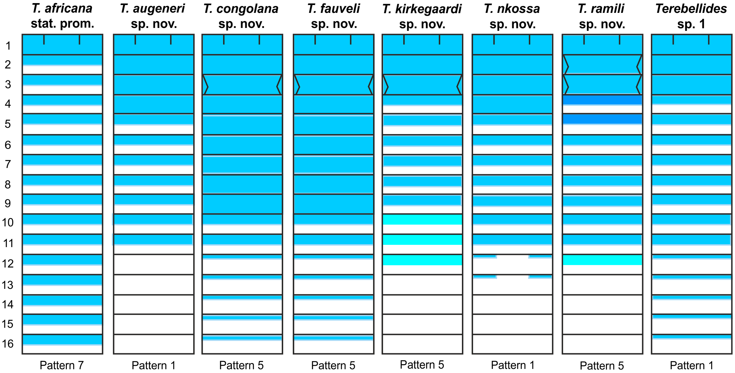
Methyl green staining pattern. Not determined; only one complete specimen available but ventrally contract- ed.
Variability. Only one complete specimen (NHMD-231448), 21 mm long, 1.5 mm wide, with 30 abdominal chaetigers. Specimen NHMD-636926 similar to holotype, with a conspicuous fan-shaped antero-dorsal expansion, branchiae with only ventral posterior lobes, very thin and with about 25 lamellae each, and nephridial openings in TN4−5. One specimen from NHMD-231448 with a scar caused by a copepod attachment near to TC1 notopodium; another complete specimen from same vial (with about 30 abdominal segments) with neuropodia as erect pinnules and about 25–30 uncini per torus having uncini with four teeth above main fang surmounted by one/two smaller ones and an upper crest of smaller teeth ( Fig. 27 View FIGURE 27 C–D). Pygidium blunt, as funnel-like depression. Specimens from NHMD-231437 badly preserved, but characteristically long, with abundant notochaetae on last half of thorax and branchiae with terminal filament in posterior ventral lobes and lamellae border conspicuously sinuous, similar to holotype.
Type locality. Equatorial Guinea; 260–650 m depth (Table 1) .
Distribution and bathymetry. From Equatorial Guinea to South Angola in SW Africa; 235–650 m depth ( Fig. 11C View FIGURE 11 ; Table 1).
Etymology. The epithet refers to the very long and numerous chaetae present in thoracic notopodia.
Remarks. The conspicuous, long thoracic notochaetae are the most distinctive character of T. longiseta sp. nov. Other species with long notochaetae are Terebellides klemani Kinberg, 1867 ( Brazil) and Terebellides irinae Gagaev, 2009 (Arctic Basin), but they are thinner and fewer, 2–4 in T. klemani , and 5–10 in T. irinae . In addition, T. klemani bears TC1 notochaetae shorter than those of following chaetigers; the branchiae of T. irinae lack anterior branchial lobes and posterior lobes originate from a thin and long stalk and are free along their entire length, the ventral being much shorter than the dorsal (see Gagaev, 2009: Fig. a–b).
Terebellides gentili Lavesque, Hutchings, Daffe, Nygren & Londoño-Mesa, 2019 also bears conspicuously long notochaetae (not explicitly mentioned in the description but see Lavesque et al., 2019: Fig. 10A View FIGURE 10 ). Nevertheless, this species clearly differs from T. longiseta sp. nov. in having fewer and thinner notochaetae and conspicuous papillar projections on the outer edge of the branchial lamellae, that are absent in T. longiseta sp. nov.
Terebellides yangi resembles T. longiseta sp. nov. in having greatly developed anterior branchial lobes but differs in length and number of thoracic notochaetae (far more numerous and longer in the African species).
On the other hand, several African non-type specimens identified as T. jorgeni may correspond to T. longiseta sp. nov. (see note below). One specimen from St. 133 showed a scar due to a parasitic copepod, similar to those reported for specimens of T. shetlandica by Parapar et al. (2016c). This was the only parasitized specimen observed across all material examined in this work.


No known copyright restrictions apply. See Agosti, D., Egloff, W., 2009. Taxonomic information exchange and copyright: the Plazi approach. BMC Research Notes 2009, 2:53 for further explanation.